Medications:
Donepezil 10 mg
Lisinopril 5 mg
Allergies:
NKA
Past Medical History:
Alzheimer’s dementia
HTN
Hearing impairment
Tobacco: 1 ppd smoker
Alcohol: none
Physical Exam:
VS: BP 101/45 (63)
HR 119
RR 32
Oxygen saturation: 99% on room air
General: moaning in pain, but able to respond to simple questions
Neuro: grossly moves all 4 extremities; unable to cooperate with sensory exam
HEENT: soot covering face, eyebrows, nosehairs singed
CV: tachycardic, regular, and without murmur
Pulm: stridorous breathing, coarse breath sounds bilaterally
Extremities: circumferential burn on right upper extremity
Otherwise insignificant
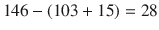
Labs: Na 146 K 4.5 Cl 103 HCO3 15 BUN 31 Cr 1.09, ABG pH 7.21 pCO2 31 pO2 67 Hg 15 mg/dL Otherwise insignificant
- 1.
How are burns classified?
Burns are classically described according to the skin layer involvement; namely, as first, second, or third degree burns. First-degree burns only involve the epidermis, are erythematous without blistering, and are painful. Second-degree burns involve the dermis; they are painful and are characterized by blistering and edema in addition to erythema. Third-degree burns involve all three layers of skin—epidermis, dermis, and subcutaneous tissue—and appear white or charred and indurated. These burns are painless as the nociceptive nerve endings have also been burned away. A less commonly described fourth-degree burn is a burn that involves not only the three layers of skin but also the destruction of fascia, muscle, and bone. First- and second-degree burns are also referred to as partial-thickness burns while third- and fourth-degree burns are full-thickness [1].
- 2.
How is the extent of burns described? Are pediatric burns measured in the same way?
The management and prognosis of the burn patient is largely dictated by the extent of the burn, termed the Total Burn Surface Area (TBSA). TBSA is approximated using one of the several tools. The palmar surface area technique uses the principle that the surface area of the patient’s palm (including fingers) represents approximately 1% of the total body surface area. This technique is reasonably accurate for small burns, or in calculating the uninjured area in very large burns and subtracting to get the large TBSA. The most common technique, the Wallace Rule-of-Nines, describes body part surface areas as multiples of 9 to approximate TBSA (see Fig. 60.1). This technique is easy to use and remember and is a good estimate of medium and large burns in adults; however, it is not accurate in children, who have a proportionally larger head and trunk surface area compared to extremities. The most accurate technique is use of a Lund Browder chart, which takes into account the age of the patient and involves a more detailed calculation of burn surface area [2, 3].

- 3.
Does this patient have a major burn?
The American Burn Association classifies a burn injury as a major burn requiring transfer to a burn unit if it includes any of the following criteria:
Partial thickness (first or second degree) of more than 25% TBSA in adults (20% at extremes of age)
Full thickness (third or fourth degree) of more than 10% TBSA
Burns (any TBSA) of sensitive areas: face, hands, feet, perineum
Inhalation burn injury
Chemical burns
Electrical burns
Burns in patients with serious coexisting medical conditions.
Therefore, yes, this patient has a major burn as described by at least 2 criteria, >10% full-thickness burns and likely inhalational injury [4].
- 4.
What is the function of the skin?
The skin is the body’s first defense against the elements. It protects our vital organs from physical damage, maintains normothermia, preserves homeostasis of fluid and electrolytes, and is involved in vitamin D metabolism. It is the first barrier to infection; a physical wall between microorganisms outside and vulnerable tissues inside. The skin is also the medium through which we perceive the world using sensations such as touch, temperature, and pain.
- 5.
What are the major physiologic derangements after a thermal injury?
The physiologic derangements seen after a thermal injury are the direct result of a disruption in the skin functions described above. Patients experience profound heat loss and the resulting hypothermia can result in cardiac arrhythmias, increased oxygen demand from shivering, and coagulopathy. Large insensible fluid losses due to evaporation result in fluid and electrolyte disturbances. Loss of skin surface area leaves these patients exquisitely vulnerable to infection. Damage to nerve endings in the skin makes burn pain severe and difficult to treat.
Most importantly, the injury triggers a large inflammatory response. It is not due to the tissue directly killed by thermal injury but by the surrounding tissue which is damaged but not dead. This damaged tissue produces local edema and inflammation, which then drives a systemic inflammatory response. It is this sublethally injured tissue that not only causes the problems but is also most at risk of further injury or death from poor perfusion or infection.
Local inflammatory mediators such as histamine, bradykinin, and prostaglandins, along with systemic mediators such as interleukins, TNF, nitric oxide, and endotoxin result in increased tissue permeability. Subsequently, a large amount of fluid moves from the intravascular space to the extravascular space in the wound, resulting in hemoconcentration and intravascular hypovolemia, even in the setting of extravascular volume overload. Intravascular volume depletion triggers a profound anti-diuretic hormone response resulting in low urine output or even anuria. This systemic inflammatory response syndrome (SIRS) leads to multiple organ failure, protein catabolism, and sepsis [5].
- 6.
Discuss the differences between a thermal burn, chemical burn, and electrical burn.
A thermal burn is caused by extremes of temperature causing destruction of the skin layers. This can be due to extreme heat or extreme cold; cold burn injury (frostbite) has its own burn classification system yet can also be associated with SIRS.
A chemical burn is caused by exposure to a corrosive substance such as strong acid, strong base, or a vesicant. They do not require heat and are sometimes not evident until several hours to days after the exposure. They have a similar classification to thermal burns, but treatment is further specialized depending on the offending agent. An additional consideration is ensuring proper protection of the staff caring for the patient to limit further exposure.
An electrical current transmitted through the body causes an electrical burn. Often the only superficial sign of electrical injury is local burn at the contact point; for example, burns on the hands where they held high voltage wire. However, the majority injury lies below the surface where electrical energy is converted to thermal energy as the current encounters resistance of various tissues in its path. In addition, damage to the heart makes it vulnerable to malignant arrhythmias [3].
- 7.
What are the effects of burn injuries to the respiratory system?
There are several different ways in which thermal injury can affect the respiratory system. Direct injury to the upper airway can quickly lead to complete airway obstruction. Even less severe injury can result in local swelling that can obstruct the airway. Inhalational injury—breathing in smoke, steam, or other noxious products of combustion—can cause both thermal and chemical damage to the smaller airways, causing significant shunting and hypoxemia. Furthermore, smoke inhalation can lead to carbon monoxide poisoning, cyanide poisoning, and other toxin exposures which vary depending on products of combustion. This can directly damage pulmonary tissues as well as decrease oxygen delivery.
Finally, the increased vascular permeability of SIRS predisposes these patients to Adult Respiratory Distress Syndrome (ARDS), which can be profound and difficult to treat due to these patients’ high fluid requirements.
- 8.
What are the cardiovascular effects of a burn injury?
The initial and rapid sequestration of fluid to the burned area results in a hypovolemic shock. The drop in intravascular volume is translated to a decrease in preload and cardiac output drops. As a result, catecholamine release triggers vasoconstriction to preserve central circulation, but this comes at a cost of ischemia to other organs such as brain, bowel, and kidneys. In addition, acidosis from carbon monoxide poisoning or hypoperfusion can directly depress cardiac function. Furthermore, hypothermia and electrolyte abnormalities from fluid shifts can predispose the heart to ectopy [5, 6].
- 9.
What are predictors of mortality in burn patients? Discuss the prognosis of this patient.
Mortality in burn patients is directly related to the TBSA. In adults, age also directly correlates with mortality. In all patients, the presence of inhalational injury dramatically increases mortality, as do coexisting medical conditions.
Our patient is 71 years old with 35% TBSA and inhalational injury. Furthermore, she may have underlying COPD resulting from her heavy tobacco use. She also appears to have some degree of renal impairment as a Cr 1.09 is inappropriately high for someone of her age. Therefore, her mortality from this burn injury would be quite high. Burn centers have long used a Baux score, defined as age + TBSA to gauge mortality from an injury. Injuries with Baux scores greater than 140 were thought to be unsurvivable. However, treatment of these injuries has improved over the years and many advanced burn centers now associate a Baux score of 140 with about 50% mortality [7]. A modified Baux score has been developed which takes into account the presence of inhalational injury, which adds about 17 years (or 17% TBSA) to the Baux score. Therefore our patient’s Baux score would be
 which is survivable by definition, but would be with a relatively high mortality [8].
- 10.
What is the most common cause of death for the burn patient?
The most common early cause of death is asphyxiation from airway obstruction due to smoke inhalation. These patients usually do not survive to be admitted to the hospital. After this early period, the most common cause of death is sepsis; in developing countries hypovolemic shock remains an important cause of mortality [8, 9].
- 11.
How will you resuscitate this patient? What fluids should you use?
The current standard of care for resuscitation uses the Parkland formula to guide fluid administration:
For the first 24 h, 4 ml/kg/% TBSA of lactated Ringer’s solution should be administered; half of the total solution given in the first 8 h, the other half given over the next 16 h. Additional crystalloid given as needed to maintain urine output at 0.5–1.0 ml/kg/hr. For the next 24 h, 5% dextrose in water can be given to maintain serum sodium in normal range (135–145 mEq/L) and colloid may be given at a range of 0.3 ml/kg/% TBSA for patients with 30–50% burn. Urine output should still be maintained between 0.5 and 1 ml/kg/hr.
Vasopressors and inotropes to maintain blood pressure should be used very judiciously; it is usually more fluid that is required, not pharmacologic agents. Additionally, diuretics such as furosemide should not be used to maintain urine output during the initial fluid resuscitation phase at risk of further damage to renal function.
For our patient at 51 kg with 35% TBSA, she should receive approximately 7.2 L fluid in the first 24 h. 3.6 L should be given in the first 8 h, 1.8 L in the next 8 h, and 1.8 L in the last 8 h. Her urine output should be above 25 ml/hr at all times [10, 11].
- 12.
What are your endpoints to determine adequate resuscitation?
In burn patients, one of the best markers of adequate resuscitation is urine output. In the absence of any drugs (diuretics, mannitol) or conditions (diabetes insipidus, uncontrolled hyperglycemia) that would falsely elevate urine output, amounts greater than 0.5 ml/kg/hr indicate a patient with adequate intravascular volume.
Other markers can offer clues to a patient’s volume status but are less reliable. Tachycardia can be due to hypovolemia or pain; the absence of tachycardia may be due to chronic beta blocker therapy, not euvolemia. Blood pressure is highly variable in patients. While mean arterial pressures (MAP) less than 60 mmHg in an adult nearly always indicate under-resuscitation, MAP greater than 60 mmHg is no guarantee of adequate perfusion of vital organs. Intense peripheral vasoconstriction may produce MAPs greater than 60 mmHg, but organs distal to the vasoconstriction are at peril. Moreover, patients with longstanding hypertension may require a higher MAP to maintain adequate perfusion pressure. Measurements of CVP can be useful at the extremes of measurement, or in observing a trend, but are largely inaccurate, particularly in patients under positive pressure ventilation. Pulmonary artery catheters can measure cardiac output, stroke volume, and estimate left atrial pressure via pulmonary capillary wedge pressure to a reasonable level of accuracy, yet the risks involved in placement and management of PA catheters preclude their use in a majority of patients. Less invasive continuous cardiac output measurements, based on arterial pulse contour, can be used as well. These allow measurement of stroke volume variation, an indicator of fluid responsiveness and therefore under-resuscitation [6, 10, 11].
- 13.
What is the cause of the patient’s acidosis?
This patient has an anion gap metabolic acidosis with a low pH and low bicarbonate.
Anion gap = Na − (Cl + HCO3); normal is 8–16.
Winter’s formula show that this is a pure metabolic acidosis with appropriate respiratory compensation:
Winter’s formula: pCO2 (expected) = 1.5 (HCO3) + 8 ± 2