CHAPTER 11 BRAIN INJURY, NEUROLOGICAL AND NEUROMUSCULAR PROBLEMS
PATTERNS OF BRAIN INJURY
The brain is extremely susceptible to injury from a variety of causes, but particularly from the effects of trauma, hypoxia, and hypoperfusion. Typical causes and patterns of brain injury are shown in Table 11.1.
TABLE 11.1 Causes and patterns of brain injury
| Traumatic brain injury | Diffuse swelling Diffuse axonal injury Acute intracerebral haematoma Acute subdural haematoma Acute extradural haematoma Contusions (bruising) Chronic subdural haematoma |
| Spontaneous haemorrhage | Subarachnoid haemorrhage Intracerebral haemorrhage |
| Cerebrovascular disease (embolic) | Stroke |
| Infection | Meningitis, encephalitis, abscess |
| Hypoxic / ischaemic injury | Watershed infarction Global infarction Hypoxic encephalopathy |
| Metabolic | Encephalopathy |
KEY CONCEPTS IN BRAIN INJURY
The principles of management are therefore to:
Cerebral blood flow and autoregulation
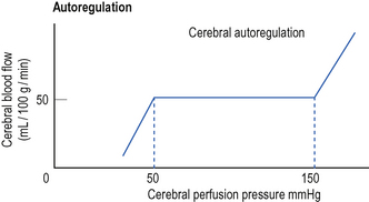
Cerebral blood flow is normally maintained at a constant level over a wide range of cerebral perfusion pressures, a phenomenon known as autoregulation (Fig. 11.1). In normotensive patients, autoregulation occurs at cerebral perfusion pressures between 50 and 150 mmHg. In previously hypertensive patients, the curve is shifted to the right and autoregulation occurs at a higher blood pressure.
Cerebral perfusion pressure
Central venous pressure is sometimes included in this equation. This is because the cranium behaves as a Starling resistor – so where the CVP exceeds the ICP, the CVP becomes the effective downstream pressure and should be used to calculate CPP.
Inadequate CPP results in inadequate cerebral blood flow and the maintenance of an adequate CPP is therefore crucial. However, there is debate about what constitutes an adequate CPP and what the target values for therapy should be. Typical target values are shown in Table 11.2.
TABLE 11.2 Typical target values for cerebral perfusion (mmHg)*
| Adults | > 60 |
| 3–12 years | > 50 |
| < 3 years | > 40 |
* There is debate about the ideal target values. Some accept lower target values.
IMMEDIATE MANAGEMENT OF TRAUMATIC BRAIN INJURY
The following notes relate to the management of traumatic brain injury. The principles apply equally well to the management of other forms of brain injury.
Airway (with cervical spine control)
The maintenance of a clear airway and the prevention of hypoxia and hypercapnia are paramount. Indications for intubation and ventilation are shown in Box 11.1.
Box 11.1 Indications for intubation and ventilation of brain-injured patient
GCS less than 8 or falling rapidly
Hypercapnia (PaCO2 > 6.5 kPa), or hypocapnia (PaCO2 < 3.0 kPa)
Inability to protect the airway
Significant facial injuries and bleeding (swelling may make intubation very difficult if delayed)
Major injuries elsewhere, especially chest injuries
Evidence of shock state (tachycardia, low BP, acidosis, etc.)
A restless patient who requires transfer to CT
Any patient with a significant brain injury requiring interhospital transfer
Intubation/ventilation of brain-injured patients
(See Practical procedures: Intubation of the trachea, p. 398.)
Establish intravenous access. Give volume loading, particularly if there is haemorrhage and other injuries. Blood, colloid or crystalloid is used as appropriate. If possible, establish direct arterial pressure monitoring.
 Check all intubation equipment, breathing circuits, ventilators and suction, etc. Monitoring should be available for BP, ECG, SaO2 and ETCO2.
 Note the baseline GCS score and pupil size for reference. The pupils are the principal clinical monitor of the brain following anaesthesia and paralysis.
 Assume there is cervical spine injury until proved otherwise. A second person should provide in-line immobilization of the neck. (It may be useful to use a bougie / airway exchange catheter / video laryngoscope to facilitate intubation without extending the neck.)
 Assume the stomach is full and perform a rapid sequence induction. Preoxygenate with 100% oxygen. Ask a trained assistant to apply cricoid pressure. Use etomidate if the BP is low, otherwise thiopentone or propofol are suitable induction agents. Suxamethonium is used to provide muscle relaxation (unless absolutely contraindicated, see p. 43).
 Opioid, e.g. fentanyl 100 μg increments or remifentanil by infusion, 0.1 μg kg−1 min−1 can be used to help block the hypertensive response to intubation.
 Ventilate to normocapnia or moderate hypocapnia (PaCO2 4–4.5 kPa). Significant hypocapnia is associated with cerebral vasoconstriction and reduced cerebral perfusion. This may potentially cause cerebral ischaemia and worsen brain injury.
 Monitor the SaO2 and ETCO2. Measure direct arterial blood pressure and blood gases as soon as possible. Maintain adequate cerebral perfusion pressure with fluids and vasoactive drugs (see below).
 Maintain sedation with benzodiazepines, propofol and opioids. During the early resuscitation and stabilization phase continue paralysis with a cardiovascularly stable non-depolarizing muscle relaxant.
Circulation
It is vital to maintain adequate cerebral perfusion pressure in brain-injured patients:
 Give colloid or normal saline to restore circulating volume. Avoid hyponatraemic fluids as these worsen outcome.
 Boluses of hypertonic saline (3–7.5%) may be valuable to maintain perfusion / circulating volume and limit cerebral oedema formation. Follow local protocols or seek advice.
Conscious level
The simplest assessment of conscious level utilizes a four-point scale:
This score is insufficiently sensitive for neurological assessment of the brain-injured patient and is only used during A&E resuscitation to give a broad indication of conscious level. Response to pain only represents a significant decrease in conscious level equivalent to a GCS score of 8 or less.
The Glasgow Coma Scale
The Glasgow Coma Scale (GCS), shown in Table 11.3, is a more comprehensive neurological assessment, which is universally used to describe conscious level and has prognostic value. It should be performed as soon as the patient is stabilized. It is repeated throughout the resuscitation process to identify any deterioration in the patient’s condition, which may suggest expanding intracerebral haematoma or brain swelling.
TABLE 11.3 Glasgow Coma Scale (GCS)
| Eye opening | Spontaneously To speech To pain None | 4 3 2 1 |
| Best verbal response | Orientated Confused Inappropriate words Incomprehensible sounds None | 5 4 3 2 1 |
| Best motor response (arms) | Obeys commands Localization to pain Normal flexion to pain Spastic flexion to pain Extension to pain None | 6 5 4 3 2 1 |
Maximum score 15. Minimum score 3. (A modified GCS is used for children under 5 years)
Reassessment and secondary survey
Having completed a primary survey and stabilized the patient, the patient should be reassessed before moving on to secondary survey. Traumatic brain injury may be an isolated injury, but this should never be assumed. The care of the brain injury must proceed alongside the continuing re-evaluation and resuscitation of the other injuries according to ATLS protocols. In particular, remember:
INDICATIONS FOR CT SCAN
CT scan should not be delayed by taking plain X-rays. Indications for CT scan are shown in Table 11.4.
TABLE 11.4 Indications for CT scan
| All patients with moderate / severe injury plus any of the following: | GCS < 13 Neurological signs Inability to assess conscious level, e.g. due to anaesthetic drugs |
| Any patient with mild injury plus any of the following: | High-risk mechanism of injury GCS < 15 for more than 2 h Skull fracture Vomiting Age > 60 years* |
* High risk patient group for occult intracranial injury
 CT normal or minimal changes only: stop sedative drugs; allow the patient to wake up and reassess neurological state.
 CT diffuse or non-operable injury (e.g. diffusely swollen brain): admit patient to ICU for further management, including monitoring of ICP.
 CT space-occupying lesion with a mass effect: requires urgent neurosurgical referral and craniotomy.
INDICATIONS FOR NEUROSURGICAL REFERRAL
The facilities available for dealing with the head-injured patient vary. Hospitals may have no CT scanner, a CT scanner but no neurosurgery, or all facilities. The decision to transfer a patient will therefore be influenced not only by the patient’s condition, but also by the local availability of resources. Indications for referral are summarized in Box 11.2.
Box 11.2 Indications for referral to neurosurgical centre
CT scan indicated but not available locally
CT scan shows intracranial haemorrhage/midline shift
CT scan suggests diffuse axonal injury
CT scan suggests raised intracranial pressure/hydrocephalus
GCS deteriorates 2 points or more
Identification of a vacant ICU bed space should not delay transfer of patients, who require an urgent CT scan or craniotomy for evacuation of a haematoma. Most neurosurgical units try to adopt an open admission policy, taking all seriously injured patients who have not had a CT scan and those who require operative intervention, regardless of the availability of ICU beds. Once appropriate interventions have been performed, any delay in finding an intensive care bed will not place the patient at further significant risk. Patients can if necessary be transferred back to the referring hospital once the need for further intervention has been excluded.
Indications for less urgent transfer include:
ICU MANAGEMENT OF TRAUMATIC BRAIN INJURY
 Use tracheal intubation and assisted ventilation to maintain adequate oxygenation and normocapnia, or mild hypocapnia (PaCO2 4–4.5 kPa).
 Maintain adequate sedation and analgesia. Paralysis is usually required in the early phases of treatment but should only be continued in unstable patients, those with raised intracranial pressure or when required to enable satisfactory ventilation.
 Nurse the patient 15–20° head-up to ensure adequate venous drainage. Avoid tight tapes to secure endotracheal tube, which may occlude jugular veins.
 Establish monitoring. Arterial blood pressure and CVP, urinary catheter, NG tube. If possible avoid internal jugular routes of cannulation (except for jugular bulb cannula). Insertion difficulties may impair cerebral venous drainage and also risk carotid injury. Use ultrasound guidance in all cases to avoid arterial puncture and other complications. The femoral route has some advantages, avoiding the need for head-down tilt during insertion.
 Give maintenance fluids as 0.9% saline (plus K+) initially. In the past maintenance fluids were restricted, but maintenance of cerebral perfusion is now considered paramount. Hyponatraemia and hyperglycaemia worsen outcome and should be avoided. Hypertonic saline (3–7.5%) have been used as boluses. Follow local protocols.
Stay updated, free articles. Join our Telegram channel

Full access? Get Clinical Tree


 Suxamethonium causes a transient rise in intracranial pressure. However, in the context of the multiply-injured patient with brain injury, securing the airway rapidly and safely is essential. Suxamethonium is usually the drug of choice.
 Endotracheal and gastric tubes should be inserted via the oral route in the fi rst instance. This is because in the presence of a base-of-skull fracture, there is a risk of these tubes entering the cranium if placed nasally. Only change to nasotracheal / nasogastric tubes once base-of-skull fractures have been excluded.


 CT scans require skilled interpretation. Do not make clinical decisions until senior experienced staff have reviewed them. Minor subarachnoid bleeding, mild cerebral oedema, early cerebral infarction, pituitary lesions and brainstem lesions are all easily missed.


