CHAPTER 10 Miscellaneous blocks
Intercostal nerve block
Preparation
• Position the patient prone, in the lateral decubitus position (with the side to be blocked uppermost) or in a sitting position. Position the patient’s arms in such a way as to pull the scapulae laterally.
• Mark the inferior margins of the ribs to be blocked just lateral to the paraspinal muscles (about 60 to 80 mm lateral to the midline for the inferior ribs and 40 to 70 mm for the superior ribs).
Technique
• Prepare the field by cleaning the skin with an antiseptic solution and positioning sterile drapes.
• Identify the target points for needle insertion – the inferior margin of the rib, just lateral to the paraspinal muscles.
• Pull the skin about 10 mm superiorly and puncture the skin with the needle parallel to the sagittal plane.
• Angle the needle 20° to 30° superiorly. The needle may be grasped with an artery forceps or a needle holder for better control (be careful not to crush the needle).
• Advance the needle until the rib is contacted (about 10 mm in the average patient, more in muscular or obese patients).
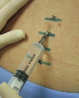
• Release the retracted skin and ‘walk’ the needle inferiorly while maintaining the superior angulation of the needle (Fig. 10.1).
• Once the inferior edge of the rib has been reached, advance the needle about 3 mm – you might feel a ‘pop’ as the fascia is penetrated. The average distance from the posterior aspect of the rib to the pleura is 8 mm, so be careful not to advance the needle too far.
• Aspirate. If no flashback of blood is obtained, inject 3 to 5 mL of local anaesthetic slowly with intermittent aspiration to rule out intravascular injection.
• Repeat the procedure with the other ribs to be blocked and one rib above and one rib below these levels.
• Local anaesthetic is rapidly absorbed after these blocks because of the high vascularity, so the toxic potential is high. Do not use more than 30 mL of bupivacaine for all the blocks combined.
Ultrasound technique
Preparation
• Position the patient prone, in the lateral decubitus position (with the side to be blocked uppermost) or in a sitting position.
• Use a linear high-frequency probe (10 to 15 MHz is ideal) and select an appropriate pre-set application.
• Identify the area to begin the scan – the appropriate ribs to be blocked plus one rib above and one rib below.
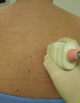
• Perform a preliminary non-sterile survey scan to identify the relevant anatomy and optimise the image by adjusting depth of field (about 20 to 30 mm), focus point, and gain (Fig. 10.2). Mark the best probe position on the skin with a pen, if required.
• Place the probe between the posterior axillary line and the paraspinal muscles in the longitudinal plane in a suitable position to obtain a transverse view of the rib to be blocked, the intercostal space and the rib below. Position the probe with the index marker superiorly so that the needle will be seen coming from right to left on the screen.
• Prepare the field by cleaning the skin with an antiseptic solution and positioning sterile drapes. Cover the probe with a sterile probe-sheath and apply sterile ultrasound gel to the appropriate area.
Technique
In-plane approach
• Identify the rib to be blocked in its transverse section, along with the rib below and the intercostal space in an area convenient to block.
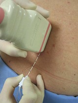
• Identify the target point for needle insertion – the superior border of the rib inferior to the one to be blocked. Position the inferior edge of the probe at the superior edge of the rib below the one to be blocked (Fig. 10.3).
• Visualise the movement of the needle as it is advanced superiorly towards the intercostal groove (the nerve itself is not visible) (Fig. 10.4A&B). Ensure that the tip of the needle is visible at all times in order to avoid penetrating the pleura.
• Once the tip of the needle is in the costal groove, aspirate. If no flashback of blood is obtained, inject 3 to 5 mL of local anaesthetic slowly with intermittent aspiration to rule out intravascular injection.
• After the procedure is complete, confirm the absence of pneumothorax on the side of the procedure with standard ultrasound techniques.
< div class='tao-gold-member'>
Only gold members can continue reading. Log In or Register to continue
Stay updated, free articles. Join our Telegram channel

Full access? Get Clinical Tree


