Figure 43.1
Inject anesthetic (Courtesy of Andrew M. Swanson MD)
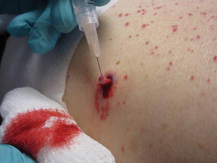
Use 1 or 2 % lidocaine with epinephrine. An allergy to procaine (Novacain) is not a contraindication to the use of lidocaine [1]. Buffer the sting with lidocaine (A 10:1 ratio of lidocaine: sodium bicarbonate 8.4 % NaHCO3) will minimize the pain (1 mL of NaHCO3 with 9 mL of lidocaine) [10]. Use a small long needle: ½ inch/gauge 27 or 26 or 5/8 inch/gauge 25. Warm the solution by gently rolling the vial between your hands. Inject perpendicular (90°) to the skin. Sensory nerve endings branch out like a tree and if the skin is penetrated at a 90° angle then the needle intersects fewer nerves [11]. The first 0.2–0.5 mL should be injected intra-dermally, to form a wheal beneath the skin. Then change the angle and slowly inject more anesthetic (See Fig. 43.2).
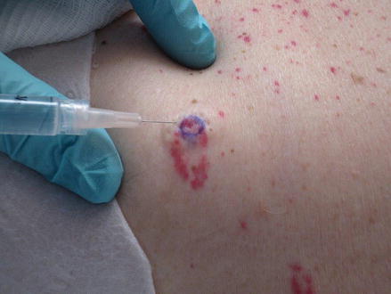
Figure 43.2
Make a wheal injecting at 45° (Courtesy of Andrew M. Swanson MD)
Keep 0.5 mL of palpable anesthetic ahead of the tip of the needle. This will anesthetize subdermal nerve endings in front of the tip of the needle [10]. Wait about 5 min for the effect of the lidocaine with epinephrine to take effect. You can use this time to fill out forms and discuss wound care with the patient. Epinephrine is absolutely contraindicated in digital and penile blocks because it may compromise blood flow [10].
Description of the Procedure
This is a clean, not sterile procedure. If the patient is at risk of infection, or immunocompromised, it should be done with sterile technique.
Non-sterile gloves
3 ml syringe filled with lidocaine with epinephrine (hemostasis is improved but may cause blanching and obscure outline of lesion)
30 g needle 0.5 or 1 inch
Punch biopsy circular blades are available in 2–8 mm diameters. A 4 mm punch tool is usually used.
Scissor
Labeled formalin containers
4–0, 5–0 or 6–0 nylon suture
Needle driver
Tissue forceps to close wound not to pick up the specimen
Clean the skin, mark with a sterile skin marker. The lesion should be outlined in case of blanching due to epinephrine with lidocaine. Stretch the skin perpendicular or 90° to the Langer’s lines. Langer’s lines are the tension lines and they usually run perpendicular to the underlying musculature but can vary within individuals (See Fig. 43.3).
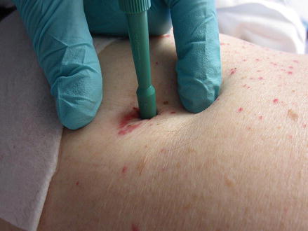
Figure 43.3
Pull skin perpendicular to Langer’s lines (Courtesy of Andrew M. Swanson MD)
To find the Langer’s lines, gently pinch the skin and look for parallel lines, hold tension in the opposite direction of the Langer’s lines when the punch is being performed. This will turn the round punch hole into a more elliptical defect that parallels the skin’s natural tension lines. This procedure will allow for a more oval shape and can be closed more easily with a suture. Place the circular blade on the skin at a 90° angle and twist back and forth until you feel a ‘give’ in the skin- you know you are in the fat when the tugging sensation is gone. Do not pull the punch tool in and out of the wound to check progress (See Fig. 43.4).
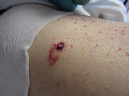
Figure 43.4
Remove the disposable punch without twisting (Courtesy of Andrew M. Swanson MD)
When you feel the ‘give’ of the fat then remove the punch and depress the skin around the defect and use the injection needle to pierce the core specimen and place in container (See Fig. 43.5).