Critical Care Medicine
Adult Critical Care
Relation between brain interstitial and systemic glucose concentrations after subarachnoid hemorrhage
Zetterling M, Hillered L, Enblad P, et al (Uppsala Univ, Sweden) J Neurosurg 115:66-74, 2011§
Evidence Ranking
• B
Expert Rating
• 2
Abstract
Conclusions
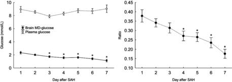
After SAH, there was a positive correlation between plasma and MD glucose concentrations with a high degree of individual variation. A gradual decline in MD glucose and the MD/plasma glucose ratio and an increase in MD pyruvate and MD lactate concentrations during the 1st week after SAH suggest a transition to a hyperglycolytic state with increased cerebral glucose consumption. The administration of insulin was related to a lowering of MD glucose and MD pyruvate, often to low levels even though plasma glucose values remained above 6 mmol/L. After SAH, the administration of insulin could impede the glucose supply of the brain (Figs 2 and 3).
Related posts:
 of change in daily step count over five years with insulin sensitivity and adiposity: population based cohort study
of Cancer: The Next Generation
Beverages and Risk of Metabolic Syndrome and Type 2 Diabetes: A meta-analysis
time and haemodynamic response after thiopental vs. propofol in the elderly: a randomized trial
monitoring of neuromuscular block over the orbicularis oris muscle in anesthetized patients receiving vecuronium
Is Better Than General Anesthesia During Endovascular Acute Stroke Interventions
of change in daily step count over five years with insulin sensitivity and adiposity: population based cohort study
of Cancer: The Next Generation
Beverages and Risk of Metabolic Syndrome and Type 2 Diabetes: A meta-analysis
time and haemodynamic response after thiopental vs. propofol in the elderly: a randomized trial
monitoring of neuromuscular block over the orbicularis oris muscle in anesthetized patients receiving vecuronium
Is Better Than General Anesthesia During Endovascular Acute Stroke Interventions
Stay updated, free articles. Join our Telegram channel

Full access? Get Clinical Tree


