Back and lower extremity pain


Back and lower extremity pain


Introduction

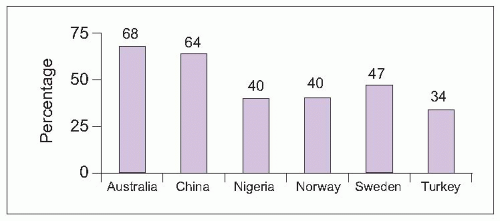
The back and lower extremities are areas commonly affected by chronic pain. Lower extremity pain may occur in conjunction with back pain (as radicular, referred, or radiating pain) or as isolated musculoskeletal or neuropathic pain. Back pain with or without associated lower extremity pain occurs frequently and is often disabling. Using data from two large, national surveys in the United States, low back pain during the preceding 3 months was endorsed by one in four adults ≥18 years old and one in three adults after age 451. The prevalence of back pain varies by region, with typically higher prevalence among developed countries (5.1)2,3,4,5,6.


5.1 World-wide 1-year prevalence of low back pain. (Based on Ihlebæk C, et al., 20062; Oksuz E, 20063; Walker BF, et al., 20044; Barrero LH, et al., 20065; Omokhodion FO, 20026.)

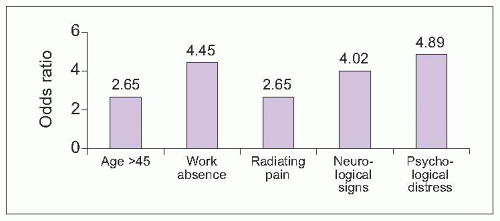
Most episodes of acute back pain resolve, with the greatest reduction in pain expected during the first 3 weeks7. Pain becomes chronic for about one in four patients. Persistent back pain can be predicted by patient demographics, physical examination, and psychological distress (5.2).


5.2 Predictors of nonrecovery from acute back pain after 3 months. Adult patients with acute low back pain for <3 weeks who contacted a primary care health provider for the first time for a back pain complaint were followed weekly for the first month and then at 3 months. Significant predictors of persistent low back pain after 3 months are shown. Work absence was significant when people lost 4 or more days during the preceding month. Neurological signs were associated with the development of chronic pain when two or more of the following were abnormal: straight leg raise, knee or ankle reflex, sensation, and strength in the thigh or foot. (Based on Grotle M, et al., 20057.)


5.3 Percentage of chronic low back pain attributable to occupation by region. (Based on Punnett L, et al., 20058.)

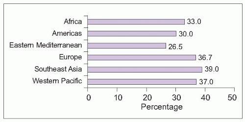
Occupation is another important risk factor for chronic back pain, with 37% of low back pain attributable worldwide to occupational factors8. Occupational contribution is highest in Europe, Southeast Asia, and the Western Pacific (
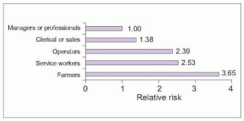
5.3). Risk varies among occupation types, with lowest risk in managerial and professional jobs and highest risk among farmers (5.4).


Assessing back and lower extremity pain

Chronic back pain is commonly caused by musculoskeletal or neurological abnormalities. Other medical conditions, including vascular, gastrointestinal, and gynecological pathology may also result in back pain (Tables 5.1, 5.2). Therefore, the physical examination must include a general medical screening, as well as abdominal and gynecologic evaluations.


Table 5.1 Nonmusculoskeletal and non-neurological causes of back pain


  • Abdominal aortic aneurysm or aorto-iliac disease


  • Abscess


  • Endometriosis or other gynecological pathology


  • Gastric or duodenal ulcer


  • Kidney disease


  • Neoplasm


  • Pancreatitis or pancreatic tumor


  • Sickle cell crisis


5.4 Relative risk of low back pain based on occupation. Compared with managers and professionals, all other occupations evaluated had a higher relative risk of low back pain based on occupation. Operators include drivers, construction and manual labour workers, plumbers, carpenters, and automobile mechanics. Service workers include nurses and other hospital workers, warehouse and stock workers, baggage handlers, waitresses, and custodians/caretakers. (Based on Punnett L, et al., 20058.)

Signs and symptoms help differentiate among common causes of back pain. Neurological symptoms or deficits suggest additional evaluations for spinal, root, or peripheral nerve abnormalities. Attention to factors that aggravate or relieve pain, localization of pain to the back or radiation to the lower extremity, and response to postural changes can help distinguish common myofascial, mechanical, inflammatory, radicular, and stenotic pain syndromes in the lumbar spine (Table 5.3).


Table 5.2 Common causes of thoracic pain


  • Myofascial pain


  • Osteoporosis with vertebral fracture


  • Metastatic neoplasm


  • Postherpetic neuralgia


Table 5.3 Differentiating common causes of chronic lumbar pain


Myofascial


Mechanical


Inflammatory


Radiculopathy


Stenosis


Pain with rest and activities


Pain worsens with activity


Pain improves with activity


Pain with rest and activities


Pain typically with walking, relieved by sitting


Nondermatomal pain*


Nonradiating pain


Nonradiating pain**


Radiates to sensory dermatome; dermatome may be numb or tingly


Pain, numbness, and cramping in both legs with walking


Muscle spasm may restrict active lumbar flexion, extension, and side bending


Restricted lumbar flexion with passive and active testing


Restricted lumbar flexion with passive and active testing


Aggravation by lumbar flexion (e.g. pain with straight leg raise); relief with lumbar extension


Aggravation with lumbar extension (e.g. walking downhill); relief with lumbar flexion (e.g. stooped gait)


* Stimulating myofascial trigger points may lead to pain radiating in nondermatomal patterns

** Alternating buttock pain may occur with sacroiliitis


Myofascial lumbar pain

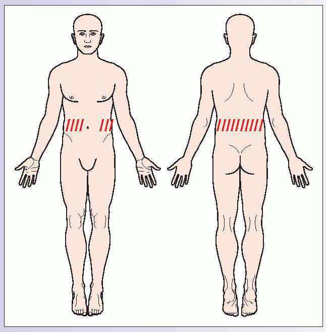
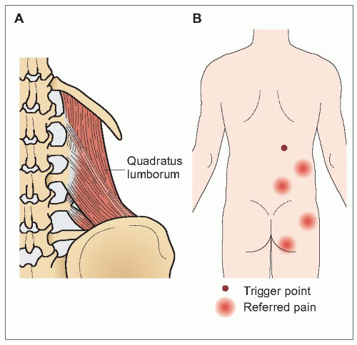
Pain of the muscles and surrounding soft tissues is termed myofascial pain. Myofascial pain is characterized by localized areas of muscle spasm and discrete points of tenderness within tight muscles, called trigger points. Trigger points are locally tender (latent trigger points) and may refer pain in predictable patterns (active trigger points) that assist in diagnosis. Myofascial pain affecting the quadratus lumborum muscle, quadratus lumborum syndrome, is one of the most common causes of low back pain. The quadratus lumborum muscles on either side of the
spine contract to cause lateral bending (5.5). Pain typically occurs in the small of the back and may be referred into the buttocks.

Piriformis syndrome is another common myofascial pain condition. The piriformis muscle connects the hip to the greater trochanter, resulting in hip stability and allowing external rotation (5.6). Pain usually occurs in the lateral buttocks with referral to the lower back and hip.


Mechanical and inflammatory lumbar pain

Restrictions of both active and passive motion in the joints in the back suggest mechanical or inflammatory pain. Pain and restrictions in joint movement in the lower back are usually caused by mechanical pain, such as degenerative arthritis. Less commonly, chronic low back pain and restricted movement may be caused by an inflammatory spondyloarthropathy. Pain and stiffness are characteristically worse later in the day with mechanical pain, aggravated by activity or exercise. Inflammatory pain and stiffness, conversely, are worse in the early morning or after resting in bed, improving with activity.


5.5 Quadratus lumborum syndrome. The quadratus lumborum muscle connects at the 12th rib, iliac crest, and lumbar vertebrae, with muscle contraction causing lateral bending of the lumbar spine (A). Quadratus lumborum trigger points occur at the waistline and may refer pain into the upper or lower buttocks (B).

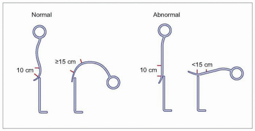
Inflammatory spondyloarthritides include ankylosing spondylitis, psoriatic arthritis, reactive arthritis (Reiter’s syndrome), and inflammatory bowel-related arthritis. While there are no specific tests for spondyloarthritides, patients often have blood tests showing elevation of inflammatory markers, like CRP, anemia of chronic disease, and the absence of rheumatoid factor. A modified Schober’s test is a nonspecific measure of reduced spine mobility (
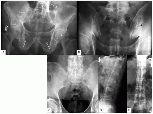
5.7). The most common spondyloarthritis is ankylosing spondylitis, which typically becomes symptomatic in young adulthood. Patients experience inflammatory changes in the spine and sacroiliac joints, with eventual fusion of the spine. While X-ray changes may be dramatic, patients typically display clinical symptoms for 5-10 years before radiographic changes are seen (5.8).


5.6 Piriformis syndrome. The piriformis muscle (1) attaches from the inner ileum (2) and sacrum (3) to the greater trochanter (4), providing hip stability and external rotation (A). Trigger points occur on the buttocks and may refer into the lower back or hip (B). The sciatic nerve (5) may become compressed beneath the piriformis muscle, resulting in leg pain. A piriformis trigger point is often performed by drawing a line from the posterior superior iliac spine to the greater trochanter and injecting immediately lateral to the midpoint of this line. The needle goes through the muscle at the site indicated by the arrow and under the muscle belly, with the injection spreading out under the muscle. 6: femoral shaft; 7: sciatic notch; 8: ischial tuberosity; 9: sacrospinous ligament; 10: posterior superior iliac spine.


5.7 Modified Schober’s test. The centre of the patient’s spine around L5, located between the posterior superior iliac spines, is marked. A mark is placed 10 cm above this point. The patient is asked to bend forward as far as possible and the distance between the two points is remeasured. This distance should now measure ≥15 cm. A new measure <15 cm suggests restricted lumbar spine flexion.


5.8 Inflammatory arthritis. Characteristic features of spondyloarthritides are shown in A (1, sacroiliac joint fusion; 2, tram track sign; 3, fluffy periosteal reaction from enthesopathy; 4, axial narrowing of hip joint with new bone formation). The point of tendon insertion into bone is called the enthesis. Inflammation of these points, called enthesitis, is common in spondyloarthritis. Ankylosing of the facet joints causes the tram track sign. Sacroiliitis is typically unilateral in psoriatic and reactive arthritis, as seen on the left in B (arrow), and bilateral in ankylosing spondylitis and inflammatory bowel-related spondyloarthritis (C). The lateral view of the spine (D) shows the square appearance of the vertebral bodies and ‘bamboo spine’ pattern characteristic of ankylosing spondylitis. The AP view (E) shows a dense central line resulting from ossification of supraspinous and interspinous ligaments, called the dagger sign.


Neurological lumbar and lower extremity pain

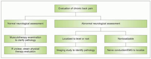
A neurological assessment of back pain patients is necessary to help differentiate musculoskeletal from neurological causes of chronic back pain (5.9). Neurological symptoms or signs suggesting radiculopathy or myelopathy, such as pain with straight leg raise testing, loss of motor strength or reflexes, and sensory loss or allodynia, warrant additional testing. Patients with motor or sensory findings suggesting a localizable spinal level or nerve root should be evaluated with a targeted imaging study, such as CT or MRI. When a particular level or root cannot be localized, nerve conduction studies with EMG should be considered.


5.9 Algorithm for evaluating chronic back pain. EMG: Electromyography.


5.10 Thoracic sensory dermatomes. Landmarks to identify thoracic dermatomes are the nipples for T4 and the umbilicus for T10.

Only gold members can continue reading. Log In or Register to continue

Stay updated, free articles. Join our Telegram channel

Jun 29, 2016 | Posted by in PAIN MEDICINE | Comments Off on Back and lower extremity pain

Full access? Get Clinical Tree

Get Clinical Tree app for offline access