Autonomic Dysfunction in Migraines


Autonomic Dysfunction in Migraines


Janne Ludwig

Thorsten Bartsch

Gunnar Wasner

Ralf Baron


A SYSTEMATIC APPROACH

Clinical symptoms like nausea, emesis, conjunctival injection, lacrimation, nasal congestion, rhinorrhea, salivation, diarrhea and polyuria have long been observed in migraine suggesting an involvement of the autonomic nervous system (ANS) (1, 2, 3, 4). Indeed, numerous studies have found a dysfunction (hypo- and hyperregulation) of the autonomic regulation in migraineurs although up to now no clear autonomic deficit has emerged from these studies which have even found contradictory results (see Tables 40-1, 40-2, 40-3, and 40-4).

In considering an involvement of the ANS many questions arise; are autonomic signs epiphenomena and secondary to the autonomic activation during migraine attacks or are they part of the pathophysiologic mechanisms involved in migraine? Is there a general dysfunction of the sympathetic nervous system (SNS) or parasympathetic nervous system (PNS) or are just some functional pathways of the systems involved? Studies revealed that the ANS and pain-relaying structures in the central nervous system (CNS) are closely coupled and often cannot be functionally separated (5, 6, 7). In addition, sympathetic pathways are functionally organized in a differentiated and specific manner so that conclusions from changes in one sympathetic pathway cannot be transferred per se to others (8,9).

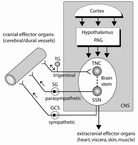
Furthermore, in view of recent experimental studies, it seems that not only the ANS, but the “neurogenic control” of the cerebral circulation is a critical issue in migraine pathophysiology. Neurogenic control not only includes the sympathetic and parasympathetic innervation, but also the afferent trigeminal and intrinsic innervation of the brain and autoregulation (see Chapter 10 and Fig. 40-1). The physiologic effects and interactions are complex and not yet fully understood. Judging by experimental evidence it seems likely that the trigeminal and parasympathetic neurogenic cerebrovascular control is not only involved in experimental pathophysiologic models, but indeed involved in the clinical setting of migraine. The clinical and experimental role of the cranial SNS in migraine is less clear.


ANATOMY AND PHYSIOLOGY

The ANS represents the involuntarily action of the nervous system; it can be divided into central and peripheral autonomic systems consisting of sympathetic and parasympathetic nerves. Sympathetic preganglionic neurons are controlled by neurons in the brainstem and hypothalamus and project from the lateral horn of the spinal cord to paravertebral ganglia of the sympathetic trunk as well as to prevertebral ganglia and synapse with the postganglionic neurons innervating effector organs. Effector organs are the adrenal medulla, smooth muscles, and glands and are involved in visceral regulation as well as cardiac and respiratory functions. Sympathetic pathways to the skin regulate blood flow and sweating by vasoconstrictor and sudomotor neurons. Noradrenaline is the predominant postganglionic transmitter of the SNS. The sympathetic innervation of the cerebral circulation originates from postganglionic sympathetic neurons accompanying the branches of the carotid artery from the cervical ganglia to mostly larger and pial cerebral vessels. Sympathetic influences on the cerebral circulation entail augmentation of the cerebral autoregulation and potential trophic effects on cerebral vessels. The involvement in migraine pathophysiology remains to be established. Other cranial sympathetic target organs include the dilator muscle of the pupil, nasal mucosa, sweat glands, and salivary glands. In addition to noradrenaline, cranial sympathetic nerves release the potent vasoconstrictive peptide neuropeptide Y (10).

Parasympathetic nerves release acetylcholine as neurotransmitter in pre- and postganglionic terminals and are involved in regulation of the heart, viscera, smooth muscles, and glands, often function antagonistically to the sympathetic innervation. Preganglionic parasympathetic
neurons synapse with postganglionic neurons in ganglia close to the effector organs.


FIGURE 40-1. Schematic view illustrating the autonomic innervation of the cranial vasculature including the trigeminoparasympathetic reflex arc as a crucial factor in migraine pathophysiology. The innervation also includes other cranial and extracranial effector organs that are not shown (see text). Nerve traffic in the trigeminal nerve may be bidirectional (afferent and antidromic). Abbreviations: CNS, central nervous system; GCS, superior cervical ganglion; PAG, periaqueductal gray; SG, sphenopalatine ganglion; SSN, superior salivatory nucleus; TG, trigeminal ganglion; TNC, trigeminal nucleus.

The parasympathetic influence on the cerebral circulation consists of a powerful vasodilatory effect, largely independent of cerebral autoregulation. Parasympathetic innervation arises from the superior salivatory nucleus and runs within cranial nerve VII through the pterygopalatine (sphenopalatine) and otic ganglia, as well as through carotid microganglia to the vessels. Other cranial target organs include the sphincter muscle of the pupil, ciliary muscle, mucosa, and salivary and lacrimal glands. Parasympathetic nerves release several, often colocalized transmitters such as vasoactive intestinal peptide, peptide histidine methionone, pituitary adenylate cyclase activating polypeptide, helodermin, and nitric oxide (11, 12, 13).


PHYSIOLOGICAL TESTS


Cerebrovascular Reactivity in Migraine

Because the ANS is involved in regulation of vascular tone, blood flow velocities (BFV) of extra- and intracranial arteries and arterial size can give information about the autonomic control of the vessels. Activity of SNS mainly results in a vasoconstriction, whereas excitation of the PNS leads to a vasodilatation (4,14).

An increased BFV refers to a small caliber of the vessel suggesting an activation of the SNS whereas an increased diameter of the vessel points towards activation of the PNS. BFV can be measured by transcranial Doppler.

A review of the literature (Table 40-1) shows that investigations of vascular reactions in migraine are ambiguous and often contradictory, probably reflecting the complex mechanisms of cerebrovascular regulation including a strong cerebral autoregulation.


Pupillometry

Pupillary diameter and responsivity can give information about the autonomic innervation of the pupil. Activation of the SNS results in dilatation of the pupil and enlarges eyelid opening, whereas blockage of the SNS causes miosis, ptosis, and enophthalmus. As in examinations of other tests reflecting autonomic functions in migraineurs different results have been obtained (Table 40-2).


Cardiovascular Reflexes

Cardiovascular reflexes can be examined using different tests such as the tilt, isometric handgrip, and cold pressure test. During the tilt test blood pressure (BP) and heart rate (HR) are monitored and patients are moved from the supine to the erect position (65 degrees). Owing to the baroreceptor reflex, HR and diastolic BP increase in healthy controls. Hypoadrenergic dysfunction—indicating sympathetic hypofunction—is characterized by a decrease of systolic (at least 20 mm Hg) and diastolic (at least 10 mm Hg) BP within 3 minutes of tilting. In contrast, sympathetic hyperfunction, as well as postural tachycardia syndrome, are characterized by a tilting-induced increase of the HR, the first being accompanied also by an increase of BP. A decrease in BP and HR owing to prolonged orthostatic exposure indicates vasovagal dysregulation.

During the cold pressor test, patients immerse one hand in icy water; under healthy conditions, this activates sympathetic muscle vasoconstrictor neurons causing an increase of the diastolic BP of at least 10 mm Hg. The same reaction occurs in the isometric handgrip test where patients press a dynamometer with 30% of their maximum voluntary power. Hand blood flow, measured with laser Doppler or venous occlusion plethysmography, indirectly refers to sympathetic vasoconstrictor innervation of the cutaneous vessels.

HR variability tests are performed during rest, deep breathing, Valsalva maneuver, and after postural changes. Pulse interval varies physiologically because of respiratory influences (sinus arrhythmia). Although both postganglionic sympathetic and parasympathetic neurons innervate the heart and influence the thresholds of excitation, HR, and atrioventricular conduction time, the inhibiting parasympathetic effect dominates under resting conditions. Therefore HR variability tests mainly reflect parasympathetic innervation of the heart.


TABLE 40-1 Cerebrovascular Reactivity in Migraineurs


Author


Methods


Point of Time


ANS Involvement in Migraineurs


Gomi et al. (42)


Retinal artery diameter


F


Sympathetic hypofunction


Iversen et al. (43)


Size of STA with ultrasound


FvA


Dilatation of STA during attack


Friberg et al. (44)


rCBF and BFV in MCA by TCD


FvA


MCA velocity on attack side lower than on nonheadache side, normal in headache-free interval; rCBF unaffected


Zwetsloot et al. (45)


BFV in MCA by TCD


FvA


No abnormalities in BFV


Thomsen et al. (46)


BFV of MCA with TCD


FvA


BFV reduced on headache side during attack


Thomsen et al. (47)


BFV of MCA with TCD during Valsalva, cold pressor test and orthostatic test


FvA


No differences in BFV during and outside of attack in physiological test; Valsalva showed mild parasympathetic hypofunction


Sliwka et al. (48)


TCD of MCA; B-waves and Mayer waves


F


Dysfunction of brain stem nuclei; impairment of sympathetic function


de Hoon et al. (49)


Ultrasound, applanation tonometry of right TA, BA


F


Right TA diameter larger; increased peripheral arterial stiffness


Thomaides et al. (50)


BFV, PI in MCA (+NO)


Nitroglycerin-induced headache in migraineurs without aura is associated with BFV changes reversible by administration of an oral triptan


Drummond et al. (51)


PA with PPT from frontotemporal region after painful stimulation of a limb


F


Limb pain triggers increases in facial blood flow


Kara et al. (52)


BFV, PI, RI in OA, PCA, CRA, VA


FvA


Retrobulbar circulation and flow hemodynamics in left VA may be altered in A and F in migraineurs without aura


Backer et al. (53)


BFV during visual stimulation before and after acupuncture in RPA and MCA with TCD


F


Abnormal cerebrovascular response in migraineurs


Boasso and Fischer (54)


TCD (BFV)


F


Elevated BFV, cerebral vasospasm in children


Silvestrini et al. (55)


Basilar and MCA reactivity evaluated with breath-holding index


F


Lower vascular reactivity in basilar artery → impairment in adaptive cerebral hemodynamic mechanism in posterior circulation


The table refers to selected studies on cerebrovascular reactivity in migraineurs indicating the diversity of results; it does not show every study performed.


Abbreviations: A, during attack; BA, brachial artery; BFV, blood flow velocities; (r)CBF, (regional) cerebral blood flow; CRA, central retinal artery; F, during headache-free interval; FvA, examination during attack versus headache-free interval; MCA, middle cerebral artery; OA, ophthalmic artery; PA, pulse amplitude; PCA, posterior ciliary artery; PI, pulsatility index; PPT, photoelectric pulse transducer; RI, resistance index; RPA, right posterior artery; STA, superficial temporal artery; TA, temporal artery; TCD, transcranial Doppler; VA, vertebral artery.


TABLE 40-2 Pupillometry in Migraineurs


Author


Methods


Point of Time


ANS Involvement in Migraineurs


Fanciullacci (56)


Pupillary diameter spontaneous, after fenfluramine and guanethidine


F


Impairment of iris sympathetic activity with adrenoceptor supersensitivity


Rubin et al. (57)


Pupillometry during cold pressure test


F


Sympathetic hypofunction


Micieli et al. (58)


Pupillary diameter


A


Either strong sympathetic activity or parasympathetic deficiency


Drummond (59)


Pupillometry, eyelid separation, thermography


FvA


Impaired ocular sympathetic outflow


Drummond (60)


Pupil diameter after cocaine, tyramine, phenylephrine eye drops


F


No evidence for adrenergic supersensitivity, but lower cervical sympathetic outflow in a subgroup


Mylius et al. (23)


Amplitude of light reflex, pupil size, latency and velocity of constriction of pupil, velocity at the end of dilatation


(A)


Parasympathetic dysfunction


The table refers to selected studies on pupillometry in migraineurs indicating the diversity of results; it does not show every study performed.


Abbreviations: A, examination during an attack; (A), within 1 week of an attack; F, examination during headache-free interval; FvA, examination during attack versus headache-free interval; MSA, muscle nerve sympathetic activity; SNS, sympathetic nervous system.


TABLE 40-3 Studies Evaluating Cardiovascular Reflexes in Migraine


Author


Methods


Point of Time


ANS Involvement in Migraineurs


Downey and Frewin (61)


Resting hand BF + cold stimulus (VOP)


F


Resting level of hand BF suggests sympathetic hypofunction, but normal reaction to cold stimulus


Steiner et al. (62)


CR


F


Sympathetic hypofunction


Gotoh et al. (63)


VM, BP, TT, Aschner test


F


Sympathetic hypofunction with denervation supersensitivity; mild parasympathetic hyperfunction in Aschner test


Drummond (64)


amplitude of pulsation of the STA, facial temperature, BP, HR, RR


F


Sympathetic hyperfunction


Cortelli (65)


TT


FvA


Impairment of SNS during migraine attack


Havanka-Kanniainen et al. (15)


HRV, TT, IWT


F


Mild parasympathetic hypofunction and sympathetic hypofunction


Havanka-Kanniainen et al. (67)


HRV (DBT, VM, TT)


F


Intact ANS in young migraine patients


Havanka-Kanniainen (66)


CR


FvA


Sympathetic hypofunction during attack


Cortelli et al. (68)


TT, HRV (DBT, VM)


FvA


Normal sympathetic function


Havanka-Kanniainen et al. (69)


PRR (DBT, VM, postural changes); IWT


F


Parasympathetic (PRR with deep breathing) and sympathetic hypofunction (VM, postural changes)


Jensen (70)


TSBF; TT


FvA


No vasomotor disturbance; normal ANS function under orthostatic condition


Kuritzky (71)


Spectral analysis of HRV


F


Imbalance in autonomic control


Havanka-Kanniainen et al. (72)


PRR (DBT, VM, orthostatic tests with TT); IWT


F


Mild parasympathetic hypofunction and sympathetic hypofunction; more severe disturbance in patients with frequent attacks


Boiardi et al. (73)


DBT, lying to standing test, postural hypotension test, sustained handgrip, HRV to VM


F


Impairment of sympathetic CR regulation in common migraine


Gomi et al. (42)


SSR


F


Sympathetic hypofunction


Mikamo et al. (74)


Orthostatic test, IWT, HRV


F


Sympathetic hypofunction


Boccuni et al. (75)


TT


F


Normal BP and HR


Cortelli et al. (76)


HR, BP, cold face test, IWT, HRV (VM, DBT; NA infusion)


F


Normal cardiac parasympathetic function, mild nonspecific sympathetic hyperactivity


Martin et al. (77)


HRV (DBT, Ewing test)


F


Sympathetic hypofunction


Pogacnik et al. (78)


VM, DBT, IWT, HRV, orthostatic test


F


Sympathetic hypofunction


Pierangeli et al. (79)


Power spectral analysis of HR, TT, VM


F


Normal function of ANS


Evers et al. (21)


SSR


F


SSR latencies are increased, amplitudes normal; authors suggest sympathetic hypofunction


Shechter et al. (80)


BP, VM, HRV, CR


F


No explicit results, rather sympathetic hyperfunction


Atasoy et al. (24)


SSR


F


SSR latencies are longer only in patients with psychiatric comorbidity, amplitudes normal


The table refers to selected studies on cardiovascular reactivity in migraineurs indicating the diversity of results; it does not show every study performed.


Abbreviations: A, examination during an attack; ANS, autonomic nervous system; BF, blood flow; BP, blood pressure; CR, cardiovascular reflexes; DBT, deep breathing test; F, examination during headache-free interval; FvA, examination during attack versus headache-free interval; HR, heart rate; HRV, heart rate variability; IWT, isometric work test; NA, noradrenaline; PRR, pulse rate reaction; RR, respiration rate; SSR, sympathetic skin response; STA, superficial temporal artery; TSBF, subcutaneous blood flow in the temporal region; TT, tilt test; VOP, venous occlusion plethysmography; VM, Valsalva maneuver.

Only gold members can continue reading. Log In or Register to continue

Stay updated, free articles. Join our Telegram channel

Jun 21, 2016 | Posted by in PAIN MEDICINE | Comments Off on Autonomic Dysfunction in Migraines

Full access? Get Clinical Tree

Get Clinical Tree app for offline access