Approach to the Patient with Lyme Disease
Lyme disease is a treatable multisystem illness caused by infection with the tick-borne spirochete Borrelia burgdorferi. The condition has become the most common vector-borne disease in the United States, with more than 40,000 cases reported to the Centers for Disease Control and Prevention in the last 10 years. In most instances, the acute infection can be readily diagnosed and effectively treated. The neurologic and musculoskeletal manifestations of later stages may be more subtle and resemble those of chronic fatigue syndrome, fibromyalgia, or depression. The nonspecificity of symptoms and shortcomings of available serologic tests lead to both underdiagnosis and overdiagnosis.
The primary physician needs to be skilled in the clinical recognition of early disseminated and late disease; clinically capable of differentiating it from other acute, subacute, and chronic neurologic and musculoskeletal conditions; cognizant of the limitations of diagnostic methods; and capable of prescribing an effective antibiotic program.
Epidemiology
The spirochete B. burgdorferi causing Lyme disease is transmitted to humans by Ixodes ticks. Nymph-stage ticks feed on humans from May through July, transmitting the spirochete in the process. Endemic areas for species of the responsible tick include the northeastern coastal states, Wisconsin and Minnesota in the Midwest, and the coast of Oregon and northern California. Outbreaks in Europe and Asia have also been reported. In the US eastern coastal regions and in the Midwest, the deer tick Ixodes dammini (scapularis) is the principal vector. More than one third of deer ticks carry the spirochete, which accounts for outbreaks of epidemic proportion. In the western United States, the Ixodes pacificus species is responsible, but the carrier rate is only 1% to 3%, and human infection is much more sporadic. The condition has also been reported in Europe and Asia.
The rising frequency of Lyme disease and its geographic spread have been linked to enlarging deer populations and concurrent suburbanization. The spirochete is transmitted horizontally to field mice, which are critical to sustaining its life cycle (deer are not, but the ticks prefer them). Human infection is a biologic dead end for the spirochete.
Deer ticks are also vectors for other important tick-transmitted diseases, which may present concurrently. Among the most important is human babesiosis, caused by the intracellular rickettsial gram-negative parasite Babesia microti, which is endemic to many of the same areas as Lyme disease. Transmission to humans is by the same deer tick, and the same field mice serve as the animal reservoir. The shared tick vector and animal reservoir increases the risk for concurrent infection. Approximately 10% of patients with active Lyme disease are concurrently infected with B. microti. Another rickettsial disease transmitted by the deer tick is human granulocytic anaplasmosis (also referred to as human granulocytic ehrlichiosis) caused by the intracellular bacterium Anaplasma phagocytophilum, which is zoonotic in the same geographic sites as babesiosis and Lyme disease. In rare instances, it too may occur concurrently with Lyme disease. The tick-borne spirochete Borrelia miyamotoi has also been associated with human granulocytic anaplasmosis.
Pathophysiology
Borrelia burgdorferi enters the bloodstream at the time of tick feeding, which usually does not occur until well after the first 24 hours of contact. After a short bloodstream phase, the spirochete organism moves out of the blood and, in seemingly trophic fashion, into the skin, synovial membranes, heart, and nervous system. After the appearance of characteristic skin lesions, there can be another period of hematogenous spread. The means by which the spirochete damages tissue is unclear; hypotheses range from direct injury to the production of antispirochetal antibodies that cross-react with tissue antigens. Patients with symptoms that persist after appropriate antibiotic therapy are suspected of having an exaggerated, sustained immune response. Such a response may account for the overlap in clinical manifestations with those of fibromyalgia and chronic fatigue syndrome, in which an immunologic pathophysiology is also suspected (see Chapters 8 and 159). The fact that patients who are positive for HLA-DR4 appear to be at increased risk for such chronic illness suggests that the severity and chronicity of disease may be related, in part, to cell surface antigens and genetic susceptibility.
Babesia microti resides predominantly in the red cells and causes mostly systemic symptoms with little localization. Infection can impair host defenses and may enhance injury caused by B. burgdorferi.
Anaplasma phagocytophilum infects white blood cells, predominantly granulocytes, and as such, it circulates in the blood stream creating the potential for transmission by blood transfusion. Severity of illness is related to degree of immunocompromise; those who are markedly immunocompromised are at greatest risk and experience higher case fatality rates.
Clinical Presentation and Course
The biting tick is usually no larger than the size of a pencil mark and often inapparent. Attachment of the tick for at least 36 hours is necessary before feeding begins and transmission of the organism takes place. A feeding nymph will appear engorged, which helps in estimating the risk of transmission when the duration of attachment is unknown. Soon after the bite, the first symptoms develop. The clinical course can be divided into three stages: (a) acute, localized disease; (b) subacute, disseminated disease; and (c) chronic disease.
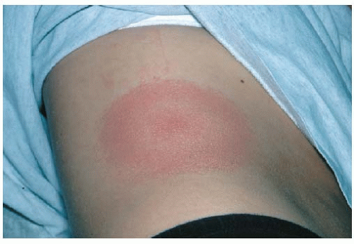 Figure 160-1 Erythema migrans. |
Stage 1: Acute Infection
Eighty percent of infected persons manifest an expanding macular erythematous rash (erythema migrans) as the first clinical manifestation of B. burgdorferi infection (Fig. 160-1). The rash usually begins as a red macule at the site of the tick bite, which spreads out to form a large, homogeneous lesion and later becomes annular with red secondary outer rings, an intense red outer border, and some clearing toward the center, although induration may be noted at the site of the bite. The lesion is large, averaging 15 cm. Minor constitutional flulike symptoms (a “summer flu”) and regional lymphadenopathy may accompany the rash. The remaining 20% of patients have flulike symptoms without a rash or no acute-stage symptoms at all. The rash starts to fade by 3 to 4 weeks. During stage 1, the immune response is minimal.
Stage 2: Hematogenous Dissemination
Hematogenous dissemination follows the acute phase within several days to a few weeks of the tick bite and leads to a host of symptoms, mostly dermatologic, musculoskeletal, and neurologic. Constitutional symptoms may be prominent, with patients complaining of generalized malaise and debilitating fatigue. Often, bouts of severe headache lasting a few hours may develop, accompanied by mild neck stiffness, as may migratory arthralgias and musculoskeletal pain.
Dermatologic Manifestations.
Dermatologic manifestations include new annular skin lesions smaller and less migratory than the initial one. Malar rash, diffuse erythema, and urticaria have also been noted.
Cardiac Involvement.
Cardiac involvement is noted in about 5% to 10% of patients, beginning several weeks into the infection. Transient heart block may be a consequence, ranging from asymptomatic first-degree atrioventricular block to complete heart block with fainting. The cardiac phase lasts 3 to 6 weeks, with the most severe forms of heart block persisting for about 1 week and not requiring pacemaker placement.
Neurologic Sequestration.
Neurologic sequestration ensues weeks to months after the initial infection, affecting 15% to 20% of untreated patients. It consists of a lymphocytic meningitis and cranial or peripheral neuropathy. The cerebrospinal fluid (CSF) shows a pleocytosis with about 100 lymphocytes/mm3, elevated protein and normal glucose levels, and antibodies to the spirochete. A mild encephalopathy may ensue and produce mood changes, somnolence, and memory disturbances. A unilateral or bilateral Bell palsy is the most common cranial nerve deficit. The peripheral neuritis presents as motor and sensory changes of the trunk or limbs in a dermatomal distribution. These neurologic manifestations can last for weeks to months.
Musculoskeletal Symptoms.
Musculoskeletal symptoms evolve into frank arthritis in up to 60% of untreated patients. The onset of arthritis is variable but averages 6 months from the time of initial infection. Characteristically, self-limited attacks of acute asymmetric monoarticular or oligoarticular arthritis develop. Pain and swelling are noted in one or a few large joints. The knee is the most common site. A joint effusion may form, composed of increased numbers of neutrophils (10,000/mm3 to 25,000/mm3). No more than three joints are usually involved in the course of the illness. Symptoms and signs last for several days to a few weeks. After an attack, the joint returns to normal.
Stage 3: Chronic Infection
After a latent period of several months and beginning 1 year after the time of the original infection, symptoms of chronic infection begin to appear. Bouts of arthritis may become more prolonged, and chronic neurologic deficits may ensue.
Skin Changes.
Most patients in the United States do not manifest skin changes in the late phases of Lyme disease, but a chronic atrophic form of acrodermatitis unique to Lyme disease has been observed in Europe.
Arthritis.
The transient form of arthritis characteristic of disseminated disease is supplanted by a more persistent one that lasts months instead of weeks. The knee remains the most common site, and the pattern continues to be oligoarticular. Joint erosion is reported but uncommon and rarely leads to permanent loss of function. In a small percentage of patients, the arthritis persists even after a full course of antibiotic therapy. An immunologic mechanism is postulated. Over the years, the frequency of arthritic episodes declines.
Neurologic Impairment.
Distal paresthesias, radicular pain, and memory loss comprise the principal neurologic manifestations of late disease, representing polyneuropathy and encephalopathy. Often, they occur concurrently. Tiredness may also be reported. In rare instances, a leukoencephalopathy with spastic paraparesis may develop. Two thirds of patients with neurologic symptoms have elevated levels of protein in the CSF, and half have Lyme antibodies in the CSF. In most patients, electrophysiologic study findings are abnormal, demonstrating evidence of axonal degeneration.
Natural History of Disease
Without treatment, 20% of patients with erythema migrans will experience spontaneous resolution and no progression of disease. Conversely, without treatment, disseminated disease develops in about 80% of patients. Attacks of oligoarthritis are common (60% to 80%) but resolve within 1 to 3 years, even without treatment. Chronic neurologic and persistent joint symptoms affect about 5% to 10% of patients. Susceptibility to late chronic disease may be genetically determined. Overall, community-based longitudinal cohort study finds that properly treated Lyme disease does not predispose to long-term disability. The view among some that Lyme disease does predispose may be a consequence of the mislabeling of incapacitated persons who do not have Lyme disease (perhaps on the basis of a false-positive serologic test).
Despite the generally favorable prognosis, a post-Lyme syndrome/chronic Lyme disease is described in up to one third of treated cases; it is characterized by persistence of fatigue, myalgias, and arthralgias for several more months after objective disease manifestations have resolved. A purported “molecular mimicry” leading to an autoimmune process has been postulated to account for some cases. All culture and polymerase chain reaction (PCR) results are negative, and prolonged courses of antibiotics (e.g., intravenous [IV] ceftriaxone 2 g/d for 30 days followed by oral doxycycline 200 mg/d for 30 days) do not improve outcomes compared to placebo. Cases of recurrent erythema migrans have been found to be the result of reinfection rather than relapse when the infecting organisms are isolated and subjected to molecular typing.
Babesiosis
The symptoms of human babesiosis include fever, chills, sweats, arthralgias, headache, and lassitude. Patients with concurrent Lyme disease and babesiosis typically manifest what appears to be a more severe and prolonged case of Lyme disease. Marked fatigue, headache, sweats, chills, nausea, conjunctivitis, emotional lability, and splenomegaly occur more frequently than in patients with Lyme disease alone. Moreover, almost half of patients remain symptomatic for more than 3 months, whereas fewer than 5% of those with Lyme disease alone remain symptomatic for that long. Similarly, patients with concurrent babesiosis and Lyme disease appear to have a more severe case of Babesia infection than would be typical.
Anaplasmosis
While concurrent infection with B. burgdorferi is rare, illness from infection with A. phagocytophilum can be severe and even life threatening if it goes undetected and untreated. Onset of symptoms is 1 to 2 weeks after the tick bite (which may be inapparent). Viral-like symptoms of headache, malaise, fever, chills, and myalgias are common and may be accompanied by gastrointestinal and pulmonary symptoms such as nausea, abdominal pain, and cough. Laboratory findings that include leukopenia, thrombocytopenia, and elevated liver function tests can provide important clues for diagnosis of the condition. Rash is not produced by the infection; its presence may suggest concurrent infection with B. burgdorferi.
Acute and Early Disseminated Stages
Lyme disease patients with acute-phase flulike symptoms, rash, and a history of tick bite may be confused with persons having Rocky Mountain spotted fever, which also is tick borne and produces an acute febrile illness with rash, musculoskeletal pain, headache, and gastrointestinal upset. However, the rash of Rocky Mountain spotted fever is different; it starts within a few days of the tick bite as an outbreak of small, blanching macules on the wrists and ankles, spreads centripetally and also to the palms and soles, and then becomes generalized and petechial. As noted previously, human babesiosis may mimic or exacerbate the early phases of Lyme disease and should be considered when a patient with a recent tick bite in an endemic area appears to have a particularly severe set of systemic Lyme symptoms that persist beyond 3 months. Anaplasmosis enters the differential diagnosis, sharing epidemiology, vector, and viral-like presentation with Lyme disease. Summertime viral illnesses round out the differential diagnosis in patients who present with flulike symptoms but without erythema migrans or a history of tick bite.
Patients with symptoms and signs of meningeal irritation require careful evaluation. Viral encephalitis is among the summer viral illnesses that may present with headache, stiff neck, and mental changes. Bacterial meningitis must also be considered. The erythema migrans and concurrent Bell palsy or radiculoneuritis of Lyme disease should help to differentiate B. burgdorferi infection from other causes of meningeal irritation.
Late Disseminated and Chronic Stages
The presence of acute episodes of oligoarthritis raises the question of gout, pseudogout, and a seronegative spondyloarthropathy (e.g., Reiter syndrome, psoriatic arthritis, ankylosing spondylitis). At the time of the initial presentation, infectious arthritis also enters into the differential diagnosis. Even early rheumatoid arthritis may present as a monoarticular or oligoarticular disease involving a large joint. The correct diagnosis can usually be made based on the associated clinical findings and the results of serologic testing and joint fluid analysis (see Chapters 145 and 146). The subtle neurologic and joint manifestations of late Lyme disease can cause considerable diagnostic confusion. Depression, fibromyalgia, and chronic fatigue syndrome have manifestations that overlap with those of Lyme disease. Clinicians encountering patients with these difficult conditions may overdiagnose Lyme disease, especially if they place too much emphasis on serologic testing and too little on symptoms and signs (see later discussion and Chapters 8, 159, and 227). Adding to the confusion is the possibility that in some instances, Lyme disease may be followed by a post-Lyme syndrome or may trigger fibromyalgia.
In general, the diagnosis of Lyme disease is made clinically, by recognizing the characteristic symptoms and signs within the epidemiologic context of the illness. Serologic testing for antibody against B. burgdorferi can supplement the clinical evaluation, particularly in persons with an intermediate pretest probability of Lyme disease, but overdiagnosis leading to overtreatment is common (see later discussion).
Early Disease
Clinical diagnosis based on history and physical examination is necessary at this stage of the illness because characteristic serologic changes will not take place for weeks. Patients are typically those who present with a tick bite, a rash, or a flulike illness. One inquires into any recent tick bite and other epidemiologic risk factors for Lyme disease (presence in an endemic area; walking in a wooded, brushy, or grassy area; and failure to take precautions against ticks). A history of tick bite is often absent (50% of persons with erythema migrans cannot recall tick exposure) and is not necessary for diagnosis. The nymph tick is very small (about 1 mm), and its bite is easily missed. After the nymph has fed, it will appear engorged (about twice its original size), which suggests that it has fed and transmitted the parasite. Diagnosis is strongly supported by the patient reporting a new migratory macular red rash on the trunk or limbs.
TABLE 160-1 Pretest Probability of Lyme Disease Based on Epidemiology and Clinical Presentation
Related posts: Screening for HIV Infection Screening for HIV Infection
 Screening for Hyperlipidemia and Associated Coronary Heart Disease Risk Factors
Evaluation of Arterial Insufficiency of the Lower Extremities
Evaluation and Prevention of Occupational and Environmental Lung Disease
Management of Chronic Obstructive Pulmonary Disease Screening for Hyperlipidemia and Associated Coronary Heart Disease Risk Factors
Evaluation of Arterial Insufficiency of the Lower Extremities
Evaluation and Prevention of Occupational and Environmental Lung Disease
Management of Chronic Obstructive Pulmonary Disease
Stay updated, free articles. Join our Telegram channel
Full access? Get Clinical Tree
 Get Clinical Tree app for offline access

|
|---|