Approach to the Patient with Dermatitis


Approach to the Patient with Dermatitis


Peter C. Schalock

Arthur J. Sober


Part 1: Atopic or Contact Dermatitis

The atopic and contact dermatitides (also referred to as eczema) are frequently encountered in medical practice, with a reported prevalence of atopic disease of up to 15% in Western industrialized countries. These conditions may be acute or chronic. The acute form is characterized by erythema, edema, vesiculation, oozing, crusting, and scaling. The chronic stage manifests excoriation, thickening, hyperpigmentation, and often lichenification. All are defined clinically by the observable changes in the skin, which reflect a common cutaneous reaction to a variety of pathogenetic stimuli. The clinical challenges for the primary care physician are to provide symptomatic relief and identify the underlying precipitant. These tasks can be difficult, often necessitating consultation with a dermatologist.


PATHOPHYSIOLOGY AND CLINICAL PRESENTATION (1, 2, 3, 4, 5, 6, 7, 8 and 9)


Clinical Presentation


Atopic Dermatitis

Atopic dermatitis is characterized by intense itching, which leads to scratching, eczematous change, and lichenification. In adults, the lesions characteristically involve the neck, wrists, area behind the ears, and antecubital and popliteal flexural areas. Nummular eczema is a variant characterized by pruritic, coin-shaped lesions on the external aspects of the extremities, the buttocks, and the posterior aspect of the trunk. The lesions may ooze, crust, and become purulent. The course varies; a few constant lesions may be present, or the number of lesions may increase gradually. The prognosis is good, with eventual clearing for most cases, although it may take years.


Contact Dermatitis

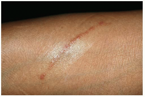
Contact dermatitis can affect any area of the body. Linear patterns are pathognomonic for plant allergens (Fig. 184-1), but almost any pattern may be seen. The distribution and the location of the rash may provide clues to the irritant or allergen,
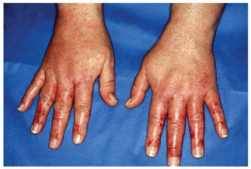
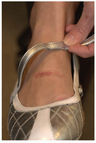
such as for a shoe (Fig. 184-2) or glove (Fig. 184-3) allergy. Patch testing can help to identify the contactant.


Figure 184-1 Linear vesicles. This pattern is characteristic of a plant allergic dermatitis—in this case, poison ivy.


Figure 184-2 Allergic dermatitis from the chromates in tanned leather in a shoe; note dermatitis under the strap.


Chronic Hand Dermatitis

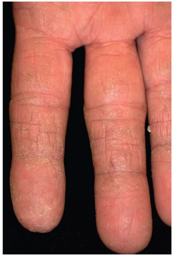
Chronic hand dermatitis presents a diagnostic and therapeutic challenge that can frustrate the most experienced dermatologist. It may be irritant in nature (e.g., “housewives’ hands”) (Fig. 184-4), pustular (psoriasis), or vesicular (pompholyx/dyshidrosis). It can occur in the context of a fungal infection with hypersensitivity reaction.


Lichen Simplex Chronicus

Regardless of the cause, chronic eczematous change may lead to lichen simplex chronicus. Itching can be intense, and the condition may be complicated by secondary infection. Lichen simplex chronicus can also result from localized neurodermatitis and present as a circumscribed plaque of thickened skin with increased markings, some scaling, and papulation. The occipital region is a common site. Lesions may also be seen on the wrists, thighs, or lower aspects of the legs. Women are more commonly affected. The prognosis is variable, but when rubbing is stopped, lesions regress.


Figure 184-3 Rubber accelerator (carba and thiuram) allergic dermatitis from latex rubber work gloves.


Figure 184-4 Chronic irritant dermatitis from frequent handwashing.


PRINCIPLES OF MANAGEMENT (1, 2 and 3,6,9, 10, 11, 12 and 13)

The management of eczema embodies the fundamental principles of dermatologic therapy: Precipitants should be eliminated, wet lesions dried, xerotic lesions hydrated, and inflammation treated with corticosteroids or calcineurin inhibitors. Resistance to treatment should be anticipated, and if basic management fails, referral to an experienced dermatologist should be considered. A search for precipitating factors is mandatory. Topical corticosteroids are frequently an important agent in treatment.


Topical Corticosteroids

Topical steroids exert anti-inflammatory, antipruritic, and antiproliferative effects. Available agents vary widely in potency, as measured by vasoconstriction assays (Table 184-1). The strongest steroid is 1,000 times more effective in blanching the skin than is the weakest. Often, differences in potency are found between generic products and those with brand names.


TABLE 184-1 Topical Corticosteroid Preparations


Group 1—Highest Potency


Betamethasone dipropionate in optimized vehicle 0.05% cream, ointment, solution (Diprolene)


Clobetasol propionate 0.05% cream, ointment, solution (Temovate)


Halobetasol propionate 0.05% cream, ointment (Ultravate)


Diflorasone diacetate 0.05% cream, ointment (Psorcon)


Group 2—High Potency


Amcinonide 0.1% ointment (Cyclocort)


Betamethasone dipropionate 0.05% ointment (Diprosone)


Desoximetasone 0.25% cream, ointment (Topicort)


Fluocinonide 0.05% cream, gel, ointment, solution (Lidex)


Halcinonide 0.1% cream, ointment (Halog)


Group 3—Medium-High Potency


Amcinonide 0.1% cream (Cyclocort)


Betamethasone dipropionate 0.05% cream (Diprosone, Maxivate)


Diflorasone diacetate 0.05% cream, ointment (Psorcon)


Group 4—Medium Potency


Desoximetasone 0.05% cream (Topicort LP)


Flurandrenolide 0.05% ointment (Cordran)


Hydrocortisone butyrate 0.1% ointment (Locoid)


Hydrocortisone valerate 0.2% ointment (Westcort)


Mometasone furoate 0.1% cream, ointment (Elocon)


Triamcinolone acetonide 0.1% ointment (Aristocort, Kenalog)


Group 5—Low Potency


Alclometasone dipropionate 0.05% cream (Aclovate)


Betamethasone valerate 0.1% cream (Valisone)


Flurandrenolide 0.05% cream (Cordran)


Fluocinolone acetonide 0.025% cream (Synalar)


Hydrocortisone butyrate 0.1% cream (Locoid)


Hydrocortisone valerate 0.2% cream (Westcort)


Triamcinolone acetonide 0.1% cream or lotion (Aristocort, Kenalog)


Group 6—Mild Potency


Desonide 0.05% cream (Tridesilon)


Fluocinolone acetonide 0.01% solution (Fluonid, Synalar)


Group 7—Lowest Potency


Dexamethasone 0.1% gel, ointment (Decadron)


Hydrocortisone 0.5%, 1%, and 2.5% cream, ointment, lotion (Hytone, Synacort, Nutracort)


It is recommended that the physician become familiar with and use one agent from each category, making the selection on the basis of cost, cosmetic acceptability, and efficacy.

Only gold members can continue reading. Log In or Register to continue

Stay updated, free articles. Join our Telegram channel

Aug 23, 2016 | Posted by in CRITICAL CARE | Comments Off on Approach to the Patient with Dermatitis

Full access? Get Clinical Tree

Get Clinical Tree app for offline access