CBP is used to temporarily perform the functions of the heart (circulation of blood) and lungs (gas exchange) during surgical procedures on the heart and great vessels. It involves a pump and oxygenator connected through an extracorporeal circuit that provides oxygenated blood flow to the systemic circulation, bypassing the heart and lungs.
A. Components of Circuit
1. Venous blood is drained from the venous cannula (inserted into the right atrium) to a venous reservoir. Vacuum assist can augment flow but is associated with hemolysis and the risk of air entrainment. Maintaining an adequate fluid level in the venous reservoir is critical to prevent air entrainment.
2. A pump (roller or centrifugal) propels the venous blood forward. Unlike roller pumps, centrifugal pumps are afterload sensitive and will decrease flow if resistance increases (e.g., with outflow obstruction). Centrifugal pumps are less traumatic to blood and do not pump air.
3. Venous blood enters a heat exchanger and an oxygenator (membrane or bubble) that adds oxygen and removes carbon dioxide by adjusting the FiO2 and sweep rate. A volatile anesthetic (e.g., isoflurane) is added to the oxygenator gas mixture. The arterialized blood passes through an arterial filter before entering the systemic circulation via an aortic cannula, which is usually inserted in the ascending aorta. The femoral artery is sometimes used for aortic cannulation for emergency or redo sternotomy situations.
4. Cardioplegic solution containing high potassium is infused into the coronary circulation to induce and maintain cardiac arrest. Cardioplegia can be infused via the aortic root or coronary ostia (antegrade) or through the coronary sinus (retrograde).
5. Left Ventricular (LV) vent removes blood that accumulates in the LV to decrease wall tension. Venting is achieved by suctioning on the same catheter placed for antegrade cardioplegia or one specifically inserted into the LV via the right pulmonary vein across the mitral valve.
6. Additional components can be connected to the circuit, including a red blood cell salvage device, an ultrafiltration unit, and an in-line unit for monitoring blood gas tensions as well as hemoglobin and electrolyte concentrations.
B. CPB pathophysiology. Contact of blood with the CPB circuit can lead to an intense systemic inflammatory response through the activation of the complement, kallikrein and coagulation cascades. A prolonged CPB time is associated with multisystem compromise including neurologic dysfunction, ARDS, coagulopathy, hepatic insufficiency, and acute kidney injury. A number of approaches have been tested to decrease inflammation in CPB including: leukocyte depletion, hemofiltration, administration of monoclonal antibodies against inflammatory mediators, and the coating of circuitry with heparin. None have shown definitive clinical benefit in humans.
C. CPB effects on pharmacokinetics. CPB results in increased volume of distribution and decreased protein binding. Acid-base shifts can affect the ionized and unionized concentrations of drugs. Decreased perfusion pressures during CPB can lead to reduced hepatic and renal clearance. Hypothermia further decreases hepatic enzyme function.
II. PREOPERATIVE ASSESSMENT FOR CARDIAC SURGERY
A. Issues pertinent to the cardiac procedure and the physiologic impact of CPB and circulatory arrest include the following:
1. Prior surgery in the chest, which technically complicates surgery
2. Evidence of aortic and cerebrovascular disease—symptomatic or documented carotid arterial disease may warrant endarterectomy beforehand. Aortic disease can affect cannulation strategies for bypass and may sometimes require concomitant repair with the cardiac operation.
3. History of bleeding, anticoagulation regimens, and prothrombotic tendencies may reveal a condition responsive to perioperative therapy.
4. A history of heparin-induced thrombocytopenia (HIT) alerts to the potential for the development of life-threatening thrombotic complications when exposed to heparin. See section on anticoagulation for cannulation and bypass for management of patients with HIT.
5. Renal insufficiency may indicate the need for intraoperative renal protective measures.
6. Patients with pulmonary disease can develop severe post-CPB pulmonary dysfunction and may benefit from preoperative antibiotics, bronchodilators, steroids, or chest physical therapy.
7. Liver dysfunction (e.g., cardiac cirrhosis) may indicate derangements in coagulation and platelet function and the need for transfusion of coagulation factors, platelets, and fibrinogen.
B. Cardiac evaluation should determine the major anatomic and physiologic characteristics of the cardiovascular system, to facilitate prediction of the likelihood of intraoperative ischemia and to determine the functional reserve of the heart.
1. Radionuclide imaging may demonstrate the regions and extent of myocardium at risk for ischemia. Radionuclide ventriculography characterizes cardiac chamber volume, ejection fraction, and right-to-left stroke volume ratios.
2. Viability studies in patients with severe LV dysfunction can help distinguish areas of myocardial necrosis from hibernation; the latter may recover after revascularization. Current modalities include nuclear imaging (SPECT or PET), dobutamine stress echocardiography, and magnetic resonance imaging (MRI).
3. Echocardiography provides an assessment of ventricular function and valve function. Regional wall motion abnormalities may reflect ischemia or prior myocardial infarction.
4. High-resolution (64-slice) computed tomographic scanning and functional MRI are noninvasive modalities to assess for coronary vascular disease. These modalities may be useful in patients who are not candidates for cardiac catheterization.
5. Cardiac catheterization remains the gold standard diagnostic test for most forms of cardiac disease.
a. Anatomic data. Coronary angiography reveals the location and extent of coronary stenoses, distal runoff, collateral flow, and coronary dominance. Significant stenosis implies a greater than 70% reduction in luminal diameter. The dominant coronary artery supplies the atrioventricular node and the posterior descending coronary artery.
TABLE 24.1 Normal Intracardiac Pressure and Oxygen Saturation
Pressure (mm Hg)
O2 Saturation (%)
Superior vena cava
—
71
Inferior vena cava
—
77
Right atrium (mean)
1-8
75
RV (systolic/diastolic)
15-30/0-8
75
PA (systolic/diastolic)
15-30/4-12
75
PA occlusion pressure (mean)
2-12
—
Left atrium (mean)
2-12
98
LV (systolic/diastolic/end diastolic)
100-140/0-8/2-12
98
Aorta (systolic/diastolic)
100-140/60-90
98
b. Functional data. Ventriculography may demonstrate wall motion abnormalities, mitral regurgitation (MR), and intracardiac shunts. LV ejection fraction is normally greater than 0.6. Impaired ventricular performance is a useful predictor of increased surgical risk.
c. Hemodynamic data are compiled from both right and left heart catheterization. Intracardiac and pulmonary vascular pressures reflect volume status, cardiac valve function, and the presence of pulmonary vascular disease (normal values are presented in Table 24.1). An elevated LV end-diastolic pressure (LVEDP) may be due to ventricular failure and dilation, volume overload (mitral or aortic insufficiency [AI]), poor compliance from ischemia or hypertrophy, or a constrictive process. The LVEDP may rise substantially in patients with coronary artery disease (CAD) after dye injection for ventriculography or coronary angiography, despite otherwise normal hemodynamic values.
d. Left-to-right intracardiac shunts are demonstrated by an arterial oxygen saturation (SaO2) “step up” in the right heart. Systemic and pulmonary flow and flow ratios can be calculated using the Fick equation.
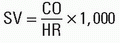
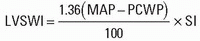
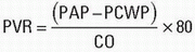
e. Cardiac output is determined by thermodilution and can be used to derive the hemodynamic indices (Table 24.2).
C. Other Studies
1. Routine laboratory studies for patients undergoing a cardiac operation usually include a complete blood count, prothrombin time (PT), activated partial thromboplastin time (aPTT), platelet count, electrolytes, blood urea nitrogen, creatinine, glucose, liver function tests, and thyroid-stimulating hormone level. A heparin-Platelet Factor 4 (PF4) antibody detection assay should be considered for patients with a low or rapidly falling platelet count associated with heparin, as these patients are at risk for developing HIT.
2. Chest radiograph and a 12-lead electrocardiogram (ECG) with a rhythm strip.
3. Pulmonary function tests may be appropriate in patients with underlying lung disease.
4. Vascular studies such as carotid duplex and vein mapping.
D. Cardiac Medications
1.β-Adrenergic antagonists, calcium channel blockers, and nitrates, including intravenous nitroglycerin, are routinely continued on schedule until the patient’s arrival in the operating room (OR).
TABLE 24.2 Ventricular Function Indices
Formula
Units
Normal Value
mL/beat
60-90
mL/beat/m2
40-60
g-m/m2/beat
45-60
g-m/m2/beat
5-10
dyne-s/cm5
900-1,500
dyne-s/cm5
50-150
BSA, body surface area; CO, cardiac output; CVP, mean central venous pressure; HR, heart rate; LVSWI, left ventricular stroke work index; MAP, mean systemic arterial pressure; PAP, mean pulmonary artery pressure; PCWP, pulmonary capillary wedge pressure; PVR, pulmonary vascular resistance; RVSWI, right ventricular stroke work index; SI, stroke index; SV, stroke volume; SVR, systemic vascular resistance.
2. Digoxin is commonly held for 24 hours preoperatively due to potential toxicity (especially in the presence of hypokalemia) and a long elimination half-life. When rate control is critical, however, as in mitral stenosis (MS), digoxin should be continued.
3. Angiotensin-converting enzyme (ACE) inhibitors, angiotensin receptor blockers (ARBs), and diuretics are usually held before surgery. Patients with significant LV dysfunction are prone to vasodilatory shock when they receive an ACE inhibitor preoperatively.
4. Antiarrhythmics are generally continued until the time of surgery.
5. Aspirin has a positive effect on graft patency and should be continued in most patients who have significant CAD. Bleeding related solely to aspirin therapy can be overcome with platelet transfusion provided the drug has been cleared from the circulation. Patients with cardiovascular pathology may be receiving multiple antiplatelet agents that may or may not be rapidly reversible (Table 24.3). Clopidogrel or prasugrel is stopped 5 to 7 days prior to surgery. If a bare-metal stent was placed within the past month or a drug-eluting stent within the last year, clopidogrel is often continued up until the time of surgery. Short-acting IIb/IIIa inhibitors should be stopped 4 hours preoperatively.
6. Warfarin is held 3 to 5 days preoperatively to allow normalization of the International Normalization Ratio (INR). Intravenous vitamin K (5 to 10 mg) or 2 to 4 units of fresh-frozen plasma (FFP) may be used emergently to correct coagulopathy. However, FFP will only transiently correct a warfarin-induced coagulopathy due to the relatively longer half-life of warfarin compared with the vitamin K-dependent cofactors ( factors II, VII, IX, and X), thus putting the patient at risk for rebound coagulopathy. Oral anticoagulants such as dabigatran, rivaroxaban, and apixaban should be stopped 5 days before cardiac surgery. Unlike warfarin, their actions cannot be reversed by the administration of vitamin K or FFP. Emergent reversal requires the administration of four-component prothrombin complex concentrates. In the case of dabigatran, hemodialysis may be used. Rivaroxaban and apixaban are too highly protein bound to be removed by dialysis.
TABLE 24.3 Antiplatelet Agents
Drug
Inhibits
Half-life
Duration
Reversible
Methods to Restore Function
Aspirin
Cyclooxygenase
15-20 min
7 d
No
Platelet transfusion
Abciximab (Reopro)
Glycoprotein IIb/IIIa receptor
30 min
48 h
Partially
Platelet transfusion
Eptifibatide (Integrilin)
Glycoprotein IIb/IIIa receptor
2.5 h
4-8 h
Yes
Delay surgery 2 h after stopping drug
Tirofiban (Aggrastat)
Glycoprotein IIb/IIIa receptor
1.5-3 h
4-8 h
Yes
Discontinue drug as soon as possible before surgery
a Dipyridamole is available in a formulation with aspirin (Aggrenox).
b Includes garlic, ginkgo, ginseng, ginger, feverfew, fish oil, and dong quai.
7. Heparin infusions initiated for unstable angina or left main CAD are continued preoperatively. The anticoagulant effects of unfractionated heparin are acutely reversible with IV protamine administration. In contrast, the anticoagulant effects of low molecular weight heparin (LMWH) preparations are not fully reversible with protamine. LMWH has been associated with increased perioperative hemorrhage in cardiac surgical patients.
a. Continuous ECG display of both leads II and V5 with ST segment trend analysis
b. Temperature monitoring includes that of the nasopharynx (reflective of the core), the blood temperature (measured from pulmonary artery [PA] catheter), and bladder or rectal temperature, which represents the average body temperature.
2. Central venous and PA pressures
a. Patients with normal ventricular function can be effectively managed with either central venous pressure (CVP) monitoring with or without transesophageal echocardiography (TEE) or a PA catheter.
b. Pacing PA catheters provide pacing capability. They can be used to maintain a suitably high heart rate in patients with regurgitant valvular lesions (AI and MR) management of a variety of valvular lesions (AI and MR) and intraoperative conduction disorders. They can also be used during procedures in patients with prior sternotomies during which rapid access for epicardial pacing may not be possible. They are also used for rapid ventricular pacing or backup pacing for transcatheter aortic valve replacement (TAVR). Mixed venous oxygen saturation (SmvO2) monitoring is available continuously with PA catheters specially equipped with a fiberoptic-linked oximeter. A decrease in SmvO2 is the result of decreased cardiac output, decreased hemoglobin, increased oxygen consumption, or decreased SaO2.
3. Intraoperative TEE is a useful tool to provide real-time information on cardiac anatomy and functional status, which can inform surgical and anesthetic decision making. The Society of Cardiovascular Anesthesiologists/American Society of Echocardiography TEE guidelines recommend the use of intraoperative in all open heart (e.g., valvular procedure) and thoracic aortic surgical procedures and to be considered in coronary artery bypass grafting (CABG) surgery. TEE may also be used to guide management during catheter-based intracardiac procedures (e.g., TAVR and MitraClips).
a. The routine examination consists of 20 standard views. The probe is advanced into the esophagus (upper and mid-esophageal views) and then into the stomach for transgastric views.
b. Application of intraoperative TEE includes assessment of global and regional LV and RV function, chamber sizes and valvular anatomy and function. TEE is very sensitive for detecting ischemia. Upon termination of CPB, it can be used to assess ventricular function, the presence of intracardiac air, and the presence of paravalvular leaks.
c. Absolute contraindications to TEE are the presence of an esophageal stricture, tracheoesophageal fistula, and a history of esophageal surgery and esophageal trauma. TEE must be used cautiously in patients with esophageal varices and altered anatomy (e.g., from gastric bypass surgery) and in those who have had radiation therapy to the neck and mediastinum. The incidence of severe complications such as esophageal perforation is on the order of 0.1%.
d. 3-Dimensional TEE can provide visualization of complex valvular features.
4. Neurologic monitors such as transcranial Doppler, multichannel electroencephalography, and near-infrared spectroscopy (NIRS) may improve neurologic outcome by alerting the clinician to perfusion imbalances during CPB. BIS monitor is a useful guide to titrate anesthetic agents for cardiac surgical patients being considered for early extubation.
B. Preinduction
1. Peripheral venous access is established. In adults, one large-bore (14- or 16-gauge) peripheral intravenous (IV) catheter is sufficient. If excessive bleeding is expected (e.g., a redo operation or a patient with a preexisting coagulopathy), a second volume line will facilitate blood product administration.
2. Sedation and analgesia are warranted in almost all cardiac surgical patients. Combinations of benzodiazepines and opioids provide excellent amnesia and analgesia for preinduction catheter insertion, with an acceptable degree of cardiorespiratory depression in all but the most debilitated patients. Patients with severe aortic stenosis (AS), MS, pulmonary hypertension, or left main CAD may not tolerate even small degrees of hypotension and respiratory depression; minimal or no premedication is administered until the patient arrives in the OR.
3. Arterial cannulation is performed with a 20-gauge catheter.
a. Left radial or femoral artery cannulation is used for systemic blood pressure monitoring when right axillary artery cannulation is planned by the surgeon for antegrade cerebral protection during DHCA.
b. Cannulation distal to a previous brachial artery cutdown site should be avoided. Pressure gradients may occur across arteriotomies, especially during and after CPB.
c. If blood pressure measurements are asymmetric, the arterial catheter should be placed on the side with the higher value.
d. Be certain to note whether the surgeon will use the radial artery for CABG surgery.
e. Femoral artery cannulation is a safe and reliable alternative to radial artery cannulation. Preoperative femoral artery cannulation in patients with severe CAD and poor LV function provides a site for postoperative intra-aortic balloon pump (IABP) insertion, if it becomes necessary. Brachial and axillary artery cannulations are third and fourth choices.
f. Intra-aortic balloon central lumen pressure can be transduced as a monitor of central arterial pressure.
4. Central venous access may be established before or after induction.
5. A defibrillator and an external pacemaker generator must be available. If the patient has a permanent pacemaker or implantable cardioverter defibrillator (ICD), a magnet should be available.
6. Typed and cross-matched packed red blood cells (2 to 4 units) must be present and checked.
7. Baseline hemodynamics values, including cardiac output, are recorded.
8. Readily available medications should include heparin, calcium chloride, lidocaine, amiodarone, inotropes, vasopressors, vasodilators, and nitroglycerin. Protamine should never be drawn up until the patient is safely separated from CPB.
IV. INDUCTION
A. Induction is a critical time in the anesthetic management of the patient. A surgeon should be available, and the CPB pump should be ready in the event of severe hemodynamic instability during induction. A systematic and gradual induction involves minimizing the degree of cardiovascular depression while maintaining an adequate anesthetic depth.
1. Agents useful in the induction and maintenance of anesthesia in the cardiac surgical patient include the following:
a. IV opioids produce various degrees of vasodilation and bradycardia without significant myocardial depression. Fentanyl (50 to 100 µg/kg) or sufentanil (10 to 20 µg/kg) can be used as both the induction and primary maintenance agents. Alternatively, a smaller induction bolus ( fentanyl 25 to 50 µg/kg) may be supplemented with a continuous opioid infusion. Alternatively, even lower doses ( fentanyl 10 to 25 µg/kg, or sufentanil 1 to 5 µg/kg) may be used in conjunction with other central nervous system depressants as part of a balanced technique.
b. Sedative hypnotics and amnestics, including benzodiazepines, propofol, and etomidate, may be useful as coinduction agents in particular situations. Of these drugs, etomidate causes the least myocardial depression.
c. Volatile inhalation anesthetics are useful supplementary agents, especially in the treatment of hypertension.
d. Muscle relaxants with minimal cardiovascular effects are commonly chosen (e.g., vecuronium, cisatracurium, and rocuronium). Pretreatment with a “priming dose” and early relaxant administration help to counteract chest wall rigidity often encountered during opioid-based inductions. Succinylcholine is used for rapid sequence inductions for patients with reflux or a full stomach. The parasympatholytic actions of pancuronium can counteract the bradycardic effects of opioids.
2. Specific considerations for valvular heart disease (see also Chapter 2).
a. Aortic stenosis (AS). Physiologic goals include the maintenance of adequate intravascular volume, sinus rhythm, contractility and systemic vascular resistance (SVR) and the avoidance of tachycardia. Patients with AS typically have a hypertrophied and noncompliant ventricle and require higher filling pressures (LVEDP of 20 to 30 mm Hg). Anesthetic agents that reduce vascular tone or myocardial contractility should be avoided. An infusion of phenylephrine can be started 1 to 2 minutes before induction to decrease the risk of developing significant hypotension associated with induction. Dysrhythmias must be treated aggressively.
b. Aortic insufficiency (AI). Physiologic goals include the maintenance of adequate intravascular volume and adequate contractility. Bradycardia and increases in SVR must be avoided. Patients with AI are often highly dependent on endogenous sympathetic tone. Patients with coexisting CAD may decompensate with significant bradycardia due to very low diastolic perfusion pressure. A means for rapid pacing should be available.
c. Mitral stenosis (MS). Hemodynamic goals mandate the maintenance of a slow rhythm, preferably sinus, and adequate intravascular volume, contractility, and SVR. Patients with severe MS and elevated pulmonary vascular resistance (PVR) are challenging to induce. Elevated PVR, often secondary to hypoventilation or positive endexpiratory pressure (PEEP), must be avoided. Atrial fibrillation with rapid ventricular response must be treated aggressively, such as with immediate cardioversion.
d. Mitral regurgitation (MR). Physiologic goals include the maintenance of adequate intravascular volume, myocardial contractility, a normal or elevated heart rate, and a reduction of systemic vascular tone. Increased SVR should be avoided. Anesthesia-induced decreases in SVR are usually well tolerated.
e. In patients with mixed valvular lesions, the most hemodynamically significant lesion will dominate the management goals. The addition of CAD to mixed valvular lesions makes planning even more complex (e.g., AS with AI and CAD). In all situations, determine the three most likely problems that could occur during induction and plan the management for each.
3. Specific considerations for emergent inductions
a. Pulmonary embolus. Induction of general anesthesia and the institution of positive-pressure ventilation can precipitate cardiovascular collapse. It is prudent to prepare and drape the unstable patient prior to induction. In patients with compromised RV function, cannulation of the femoral vessels under local anesthesia should be performed prior to induction to allow for the emergent institution of CPB if needed.
b. Pericardial tamponade. Similar concerns are present for patients with pericardial tamponade. Adequate volume administration is essential. Starting an inotropic agent and a vasopressor before induction may be helpful. A rapid sternotomy may be required if hemodynamic collapse occurs with anesthesia induction. If possible, the pericardial effusion should be drained under local anesthesia prior to induction.
c. Aortic dissection. Hypertension can precipitate aortic rupture. Packed red blood cells must be available in the OR before induction. Proximal extension of the dissection into the coronary arteries can occur leading to myocardial ischemia or tamponade.
d. Ventricular septal defect (VSD) and papillary muscle rupture after myocardial infarction. Patients may present with extreme hypotension. Rapid initiation of CPB is essential. Preinduction initiation of intraaortic balloon counterpulsation therapy is indicated in many of these patients.
B. The prebypass period is characterized by variable levels of stimulation during the preparation for CPB initiation. Stimulating periods include sternotomy and sternal retraction, pericardiotomy, and aortic cannulation.
1. Baseline PaO2, PaCO2, pH, hematocrit (Hct), and activated clotting time (ACT) should be obtained.
2. Consider autologous blood procurement in otherwise healthy patients with a starting Hct of 40% or greater. 1 to 2 units of whole blood may be drained passively into sterile blood bags and later transfused into the patient following conclusion of CPB and heparin reversal.
3. The lungs are deflated during sternotomy. Physical alterations of the chest wall can produce ECG changes (especially T-wave changes), which should be noted to avoid confusion with ischemia.
4. Left internal mammary artery dissection may cause ispilateral hemothorax and negatively affect pulmonary mechanics in patients with reduced pulmonary reserve.
5. Anticoagulation for cannulation and bypass
a. Prior to the induction of anesthesia, heparin at a dose of 350 units/kg should be drawn up and kept readily available in case the emergent initiation of CPB is necessary. The dose may be increased to 500 units/kg if the patient has been receiving heparin infusion or is being treated with IABP counterpulsation. The heparin should be administered through a centrally placed catheter.
b. Vasodilation often follows the heparin bolus and should be anticipated.
c. The ACT is used to monitor the degree of anticoagulation. It should be measured approximately 5 minutes after heparin administration. Baseline values are 80 to 150 seconds. Heparin treatment sufficient to prevent microthrombus formation during CPB correlates with an ACT of more than 400 seconds (at higher than 35°C). An ACT value of greater than 450 seconds is preferred, given the variability that exists in this point-of-care test. Patients on continuous IV heparin preoperatively may become relatively “heparin resistant.” If an ACT longer than 400 seconds is not achieved with the standard heparin dose, an additional 200 to 300 units/kg is administered. If this fails, antithrombin concentrate (500 to 1,000 units) or 2 to 4 units of FFP may be necessary to correct a probable antithrombin III deficiency.
d. Patients with a diagnosis of HIT type 2 (also known as HIT with thrombotic syndrome [HITTS]) require alternative anticoagulation management during CPB. The classification of HIT is determined by immune involvement. HIT type 1 is a nonimmunologic reaction of heparin with platelets that causes a mild thrombocytopenia. HIT type 2 is an immune-mediated phenomenon that activates platelets, resulting in platelet aggregation. Biochemical mediators from activated platelets can induce the generation of thrombin, leading to diffuse arterial and venous clotting. The diagnosis requires serologic and clinical evidence. Patients with a positive functional assay (serotonin release assay or platelet aggregation study), greater than 50% reduction in platelets (irrespective of starting platelet count), drop in platelet count less than 150k, or history of a thrombotic event associated with heparin use are more likely to have an adverse outcome when re-expose to heparin. Patients with a positive ELISA test in the absence of a positive functional assay or clinical symptoms have a lower likelihood of having an adverse reaction to heparin.
e. For patients with HIT type 2 or HITTS, alternatives to standard heparin treatment exist (Table 24.4); each has significant limitations that should be discussed with the surgeon and a hematologist before use.
1. All forms of heparin are removed preoperatively. Saline is used to flush pressure transducers, and citrated saline is used to wash salvaged blood during the centrifugation process.
2. A heparin-free PA catheter is used.
3. Alternative anticoagulant regimens may be used. These include bivalirudin, or unfractionated heparin in combination with an antiplatelet agent (see Table 24.4).
TABLE 24.4 Alternative CPB Anticoagulation in Patients with HIT