Anatomic variants are variations in normal anatomy that can be misinterpreted as pathological conditions. Many anatomic variants are remnant structures from embryological development and fetal circulation, particularly in the atria. Anatomic variants are seen in multiple image planes and persist despite changes in transducer frequency, gain, compression, and depth. Ultrasound artifacts are errors in imaging most commonly due to a violation of the assumptions that are inherent in any ultrasound system. All imaging systems assume that sound travels in a straight line, travels directly back from a reflector, and travels at exactly 1540 m/s through soft tissue. Additionally, it is assumed that the ultrasound beam is very thin, reflections are entirely from structures within the main axis of the beam, and the intensity of reflections is related only to the tissue characteristics of the reflector.1 Artifacts cross known anatomic planes and boundaries and typically disappear with alternate imaging planes and when remedial actions are taken. It is vital to be familiar with the common anatomic variations and ultrasound imaging artifacts to ensure accurate echocardiographic interpretation and to avoid unnecessary interventions.2
The atria and the sinus venosus evolve in the 4th week of embryonic development. Initially, the sinus venosus receives venous blood from left and right sinus horns (Figure 3–1A and B). In time, the veins to the left sinus horn are obliterated and the remnants become the coronary sinus. The right sinus horn, on the other hand, enlarges and forms the smooth-walled part of the right atrium (RA), known as the sinus venarum. As the RA expands, the sinus venarum displaces the trabeculated tissue of the primitive RA into the periphery and into the right atrial appendage (which may have prominent pectinate muscles). Right and left venous valves mark the junction of the original sinus venarum and the primitive RA. The left venous valve disappears as it fuses with the developing atrial septum. The right venous valve of the right sinus venosus horn develops inferiorly into (1) the valve of the inferior vena cava (IVC) or the eustachian valve, which directs fetal blood flow from the IVC across the foramen ovale, and (2) the valve to the coronary sinus or the thebesian valve (Figure 3–2). Superiorly, the convergence of the smooth sinus venarum and the trabeculated RA is the crista terminalis. Concurrently, the atrial septum forms with migration of the septum primum to obliterate the ostium primum, followed by the migration of the septum secundum to cover the ostium secundum. This migration leads to the characteristic thin-walled appearance of the foramen ovale, with incomplete septation leading to the possibility of a patent foramen ovale.3


In the left atrium (LA), the smooth tissue of the pulmonary veins is incorporated into the wall of the left atrium and it displaces the primitive atrial trabeculated tissue almost entirely into the left atrial appendage. The ridge of tissue at the junction of the smooth left superior pulmonary vein and the trabeculated left atrial appendage is called the ligament of Marshall or, more colloquially, the coumadin ridge (since this structure was initially misinterpreted as a thrombus requiring anticoagulation). During the 8th week of embryonic development, the distal end of the left common cardinal vein degenerates, and the proximal portion connects via the left brachiocephalic vein to the right brachiocephalic vein, forming the superior vena cava (SVC). The left posterior cardinal vein also degenerates and the remnants of the left sinus horn, receiving venous drainage from the heart, become the coronary sinus. Failure of the left posterior cardinal vein to resorb results in a persistent left superior vena cava (PLSVC) that drains into and dilates the coronary sinus.
The anatomic variants are best classified by location, although some variant structures can appear in more than one cardiac chamber.
The crista terminalis is seen at the junction of the SVC and the RA, forming a structure that may appear to protrude longitudinally into the RA towards the IVC. This structure is often visualized in the midesophageal (ME) bicaval view and should not be mistaken for thrombus or tumor (Figure 3–3). Of note, the crista terminalis is thought to be a location where atrial tachydysrhythmias originate due to the high density of adrenergic nerve fibers, and thus may be a site for ablation therapy.4

The eustachian valve can be found in multiple views including the ME bicaval view and right ventricular (RV) inflow-outflow views (Figure 3–4). It is seen at the junction of the IVC and RA in approximately 25% of individuals and appears as an elongated, membranous, sometimes undulating structure that can extend from the IVC to the border of the fossa ovalis. Usually it is of no physiological consequence, but it can be confused with an intracardiac thrombus, cause turbulent atrial blood flow, complicate IVC cannulation, or serve as a site for endocarditis or thrombus formation.5 Occasionally it may also appear to bisect the right atrium, simulating cor triatriatum dexter, but a eustachian valve is distinguished by a lack of flow disturbance on color-flow Doppler examination.6
The thebesian valve is a structure that can be seen as a thin piece of tissue guarding the entrance to the coronary sinus in the ME four-chamber view with the probe slightly advanced towards the tricuspid valve. It can also be seen in a modified bicaval view inferior to the left atrium in the atrioventricular groove (Figure 3–5). This valve serves to prevent retrograde flow into the coronary sinus during atrial contraction and is inconsequential unless it inhibits cannulation of the coronary sinus for retrograde cardioplegia catheter placement or biventricular pacing wire advancement.7

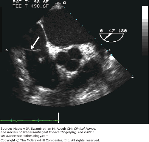
The Chiari network is a thin, mobile, membranous structure seen within the RA in multiple imaging views (Figure 3–6) that is thought to be a remnant of sinus venosus–derived structures. It is similar to but usually more extensively attached to intracardiac structures than the eustachian valve. The Chiari network is typically perforated and associated with the IVC orifice; however, the primary site of origin can vary to include the RA wall, interatrial septum, or the coronary sinus. The Chiari network moves toward the tricuspid valve during atrial contraction followed by a rapid posterior motion at the onset of ventricular systole. It has little clinical significance except that it has been associated with a patent foramen ovale, interatrial septal aneurysm, and paradoxical embolization. It is seen in 2% to 3% of all patients at autopsy and by TEE.8

The coronary sinus is best seen with slight probe advancement in the ME four-chamber view as an echolucency in the RA, just superior to the tricuspid valve (Figure 3–7). Since it courses in the atrioventricular groove superior to the mitral valve annulus,9 it can also be seen in a modified ME bicaval view as it curves around the atrium in the atrioventricular groove (see Figure 3–5), or in cross-section in the ME two-chamber view (Figure 3–8). It is a useful structure to identify in order to assist with the placement of coronary sinus catheters for retrograde cardioplegia delivery and pacing wires for biventricular pacing. Despite echocardiographic guidance, injury to the coronary sinus during these procedures is not uncommon.10 Understanding the normal anatomy of the coronary sinus is also important for identification of an inferior sinus venosus defect. The coronary sinus is normally less than 1 cm wide and approximately 3 cm long, but can dilate as a consequence of right heart volume or pressure overload.11 Coronary sinus dilation can result from atrial hypertension, tricuspid regurgitation, or a PLSVC that drains into the coronary sinus (Figure 3–9).12 A PLSVC can also be seen between the left upper pulmonary vein and the left atrial appendage in the ME four-chamber view (Figure 3–10). Diagnosis of a PLSVC is suggested by a dilated coronary sinus (> 1.1 cm) and confirmed by injection of agitated saline into a left upper extremity vein resulting in opacification of the coronary sinus as the PLSVC flow enters the coronary sinus (see Figure 3–10) and then into the RA.


The normal foramen ovale is seen best in a ME bicaval view; it appears as a thin slice of tissue bound by thicker ridges of tissue, one of which appears as a “flap.” Up to 30% of the population may have a probe patent foramen ovale (PFO), with the possibility of right to left intracardiac shunting (Figure 3–11) when right atrial pressure exceeds that of the left atrium.13 TEE evaluation of the foramen ovale should include 2D assessment for flap movement and color-flow Doppler assessment, optimized for measurement of lower velocity flow. Injection of agitated saline (a “bubble study”) along with a Valsalva maneuver is typically used to provoke right to left shunting. In such a study, the bubbles should be injected after the Valsalva maneuver produces a decrease in RA volume, and the Valsalva should be released (so as to transiently increase RA pressure over LA pressure) when the microbubbles are first seen to enter the RA. Admixture of agitated saline with small quantities of blood has been reported to improve the acoustic signal of the microbubbles. The bubble study is positive if bubbles appear in the left atrium within five cardiac cycles (Figure 3–12).


This condition may be idiopathic or may develop as the result of right heart dysfunction and elevated right-sided pressures.13 The interatrial septum is enlarged and seen to be undulating between each atrium during the cardiac cycle (Figure 3–13). An interatrial septal aneurysm is defined as constituting more than 1.5 cm of the atrial septum and extending 1.5 cm into either atrial chamber (Figure 3–14). The grading system for these aneurysms is largely based on the extent of excursion into the left and right atrium (see Appendix H). Atrial septal aneurysms have been associated with PFO and Chiari network and may predispose to thrombus formation, resulting in potential paradoxical embolism and stroke.14 Percutaneously inserted closure devices for PFOs may be efficacious in patients with paradoxical emboli.15


Lipomatous thickening of the interatrial septum is often quite striking, and may mimic an infiltrative process. This benign process creates a dumbbell-like appearance of the superior and inferior atrial septum, and is characterized by the lack of involvement of the fossa ovalis (Figure 3–15). The echogenic fat may also involve the right atrial wall, a finding that is associated with coronary artery disease and obesity.16

Trabeculations seen on echocardiographic examinations represent the muscle bundles on the endocardial surface of the heart and are more characteristic of the RA, right atrial appendage, and right ventricle than the left atrium and ventricle. Right ventricular hypertrophy may accentuate these trabeculations.
A series of parallel ridges known as pectinate muscles course across the anterior endocardial surfaces of the left and right atria, including both appendages. Pectinate muscles are more apparent in the RA than in the left atrium (Figure 3–16). Prominent pectinate muscles can be distinguished from a mass or thrombus by their movement in synchrony with cardiac tissue, whereas a thrombus is often asynchronous with cardiac motion and is associated with arrhythmias such as atrial fibrillation or low flow states such as mitral stenosis.17

The RAA is most commonly seen in a ME bicaval view where the crista terminalis separates the SVC and RAA. Occasionally, the prominent trabeculations or pectinate muscles can also be seen. The RAA can also appear as an echo-free space anterior to the ascending aorta and near the right ventricular outflow tract in the ME aortic valve long-axis view.
The atrial tissue separating the entrance of the left upper pulmonary vein (LUPV) from the left atrial appendage (LAA) commonly has multiple appearances, including a globular fatty appearance, often resembling a “Q-tip” (Figures 3–17 and 3–18). It is commonly referred to as the “warfarin” or “coumadin ridge” because it has historically been misinterpreted as a thrombus leading to treatment with anticoagulants. The ligament of Marshall is an important landmark for electrophysiological ablation procedures as it is thought to contribute to the maintenance of atrial fibrillation.18
Related posts:


Stay updated, free articles. Join our Telegram channel

Full access? Get Clinical Tree


