Fig. 11.1
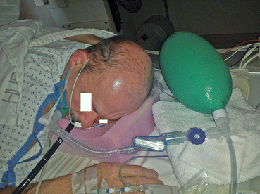
A nasal airway connected to a Mapleson C breathing system. Also seen in the picture is the sensor for SEDLine depth if sedation monitor
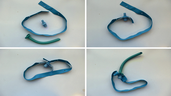
Before the nasal airway is inserted, it is also important to prepare the airway itself, so that it can be secured. There are multiple ways of achieving this. Although tape can be used, it might not be stable. The way practiced at the hospital of the university of Pennsylvania is demonstrated in Fig. 11.2. An endotracheal tube connector is obtained. They can be either purchased on their own or removed from a new endotracheal tube. Using the steps in the figure, the endotracheal tube connector and the nasal airway are held together. Typically, a 32 fr size is used for adult females and 34 fr for an adult male. Smaller airway might be appropriate for smaller size adults of both gender.


Fig. 11.2
Various steps to prepare a nasal airway before insertion and thereafter connection to a Mapelson C breathing system
After insertion of the nasal airway, it is connected to a pre-prepared Mapleson breathing system, which was used during pre-oxygenation. An oxygen flow of 6–8 l is typically used, however it can be increased to allow for the leaks in the event of attempted ventilation. It is important to maintain a sedation depth that would allow spontaneous ventilation. Such a degree of sedation would allow scope insertion including maneuvering required for advanced endoscopic procedures. Although no prospective controlled studies exist on the safety and efficacy of this technique during gastrointestinal endoscopy under deep propofol sedation, sufficient body of retrospective data is available [9–11].
In addition to providing high concentration of oxygen at the laryngeal inlet, the nasal airway might provide a degree of positive pressure ventilation by occluding the second nostril and mouth around the endoscope. However, it is by no means guaranteed to provide a good seal or allow consistent intermittent positive pressure ventilation (IPPV). In the event of desaturation due to apnea, if the initial efforts at IPPV fail, it is very important to request the endoscopist to withdraw the endoscope and initiate positive pressure ventilation.
As a result of propofol deep sedation, muscles of the jaw become relaxed and the tongue falls back to obstruct the oropharynx. This might be associated with obstruction of the nasopharyngeal air passage brought about by the soft palate falling back onto the posterior nasopharynx. Chin lift, jaw thrust and suctioning of the oropharynx are often forgotten and needs implementation early on. Any laryngospasm needs to be identified on time and appropriate measures including administration of succinylcholine instituted.
Oral Airway Connected to a Portable Mapleson Breathing System
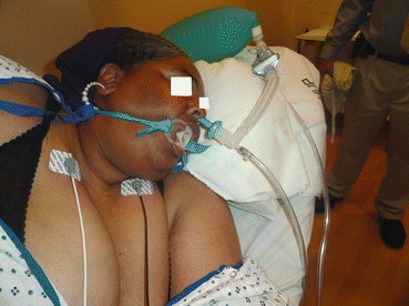
Figure 11.3 illustrates the nasal airway inserted on the side of the month to achieve similar objectives as nasal airway. Securing the trumpet is similar to the nasal airway. Once again, no randomized studies are available to confirm its safety and efficacy.


Fig. 11.3
A nasal airway in the mouth which is connected to a Mapleson C breathing system
Using either of the above techniques would limit the ability to sample the end tidal gas for a reliable display of end tidal carbon dioxide graph or number. This limitation was studied and reported [12]. It might be sensible to use alternate means of respiration like impedance pneumogram or acoustic respiratory monitoring. The limitation of the former is its inability to detect breathing against a closed glottis, while the latter is expensive and insufficiently studied.
Nasal Airway Connected to a Jet Ventilator
Supraglottic jet ventilation is occasionally employed in upper GI endoscopy. As displayed in the Fig. 11.4, the nasal airway is connected to the luer connector of jet ventilator tubing.


Fig. 11.4
A nasal airway connected to a C Mapleson breathing system. The Luer fitting on the elbow of the breathing system is connected to a hand held jet ventilator
High frequency Jet ventilation (HFJV) uses a high-pressure gas source which is applied to an open airway in short bursts. This can be achieved with a hand held device (manual jet ventilation originally described by sanders), where rate and duration of the breath are set by the user depending on the chest expansion and the saturation. An automated system that delivers oxygen or air-oxygen mixture, at respiratory rates substantially higher than physiologic (between 60 and 300 breaths/min, often termed HFJV) is another option. This allows for a nearly motionless operative field as well as frees the anesthesiologist from ventilation responsibilities during the procedure. Patients with restrictive lung disease, poor chest wall compliance, and significant obesity are poor candidates for automated jet ventilator. They can develop atelectasis during prolonged procedures due to low VTs (small tidal volume) and low mean airway pressure. Development of excessive airway pressures and pneumothorax is a limitation of manual jet ventilation.
Unlike in rigid bronchoscopy where jet ventilation is popular, its use is limited to few enthusiasts in the area of GI endoscopy. A case can be made for its use in patients undergoing Endoscopic Retrograde Cholangiopancreatography (ERCP). However, it is important to align the tip of the nasal airway with the laryngeal opening. It is also essential that the tip of the nasal airway is not obstructed as this can lead to potential air entry into the soft tissues (as pharyngeal trauma cannot be excluded either during nasal airway or endoscope insertion) with catastrophic airway occlusion. Morbidly obese is another potential group who can benefit from jet ventilation [13, 14]. There are no studies conforming its safety and efficacy in the setting of GI endoscopy.
Stay updated, free articles. Join our Telegram channel

Full access? Get Clinical Tree


