Adrenal Dysfunction


Adrenal Dysfunction


Abeer Hassoun

Sharon E. Oberfield


The pathophysiology and management of adrenal disorders that are relevant to the care of children in the ICU are reviewed in this chapter.


BIOLOGY OF ADRENAL FUNCTION


Overview

Four hundred and fifty years ago, Bartholomeo Eustacius (1) described the anatomy of the adrenal gland; shortly thereafter, the zonation of the gland and the distinction of the cortex and medulla were described. The human adrenal glands are located superior to the upper pole of each kidney and can be considered as two unique endocrine organs: the adrenal cortex and the adrenal medulla. Thomas Addison (2) defined the function of the adrenal gland. However, it was really 100 years later, in the 1950s, after performing adrenalectomies in animals, that Brown-S’equard (3) described the adrenal glands as “organs essential for life.” Pituitary control of the adrenal function was reported in the 1920s, and this was followed by the isolation of sheep adrenocorticotropic hormone (ACTH) (4). Corticotropin-releasing hormone (CRH) (the hypothalamic releasing factor) was not synthesized until 1981 (5). With respect to the adrenal glomerulosa, primary aldosteronism was described by Conn (6) in the mid1950s, and the regulation of aldosterone by angiotensin II demonstrated afterwards. It is now known that transcription factors are essential for the development of the adrenal gland: SF-1 (steroidogenic factor-1) and DAX1 (dosage-sensitive sex reversal, adrenal hypoplasia congenita, X chromosome) (7).

The adrenal cortex consists of three zones: the outermost, zona glomerulosa (15%), from which there is a gradual transition to the middle, zona fasciculata (75%), and then a clear transition to the inner most, zona reticularis (10%). The zona glomerulosa contains relatively small cells with a low cytoplasmic-to-nuclear ratio. It is the primary site of mineralocorticoid synthesis. The zona fasciculata has large cells with a high cytoplasmic-to-nuclear ratio and a large number of cytoplasmic lipid vacuoles, which are arranged in columns directed toward the center of the gland (radial cords). This zone is involved in the production of cortisol. The zona reticularis contains compact anastomosing cords of cells and an intermediate cytoplasmic-to-nuclear ratio. The zona reticularis produces predominantly androgens.

The adrenal medulla should be considered a part of the sympathetic nervous system and is composed of chromaffin cells arranged in nests and cords with sympathetic ganglion cells. In adults, epinephrine is the major catecholamine synthesized and stored in the adrenal medulla. However, this varies with age, for example, at birth norepinephrine is the major catecholamine (8).

In cord blood and in newborns, the concentrations of both cortisol and cortisone are low and about equal, 4-10 µg/100 mL of plasma (9). After 3-4 weeks of life, the cortisol concentration increases in relation to that of cortisone. There is little or no diurnal variation of glucocorticoid concentration until about 4-6 months of age (10). Cortisol secretion rates, corrected for body surface area, remain constant during childhood, puberty, and adulthood, and maintain a diurnal variation. In parallel with cortisol, ACTH peaks between 6 a.m. and 9 a.m, declines to nadir between 11 p.m. and 2 a.m., and then start to rise between 2 a.m. and 3 a.m. (11). Urinary-free cortisol excretion may be higher in perimenarcheal females (12). Aldosterone is secreted at a constant rate during infancy, childhood, and adulthood, albeit the concentrations of aldosterone in infancy tend to be relatively higher than those observed later on in childhood for specific levels of sodium (13). Adrenal androgen secretion is low during childhood. Prior to puberty with onset of adrenarche (physically noted as the onset of pubic hair), there is an increase in the secretion dehydroepiandrosterone (DHEA), dehydroepiandrosterone sulfate (DHEA-S), and androstenedione (9). At the same time, however, there is no change in ACTH concentrations in blood or of the cortisol secretion rate corrected for body size raising the issue as to what stimulates the increase in DHEA/DHEA-S secretion.


Adrenal Cortex


Hormones Produced by the Adrenal Cortex

Three main classes of hormones are produced by the adrenal cortex: glucocorticoids (e.g., cortisol), mineralocorticoids (e.g., aldosterone), and sex steroids (e.g., testosterone, DHEA, and androstenedione).

All steroid hormones are derived from the cyclopentanoperhydrophenanthrene structure that is composed of three cyclohexane rings and a single cyclopentane ring. Although adrenal steroid cells can synthesize cholesterol de novo from acetate,
80% of the cholesterol precursor for adrenal cortex hormone synthesis is provided from circulating plasma lipoproteins. Cholesterol (mainly LDL in circulation) is the precursor for all adrenal steroidogenesis (14).

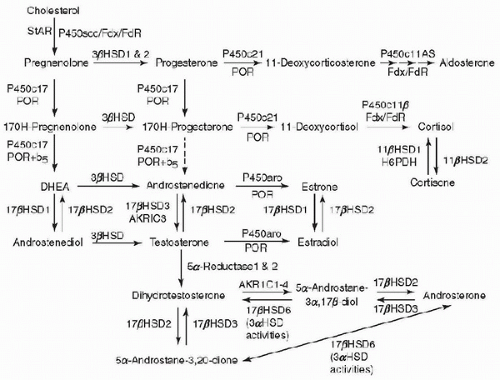
The biochemical pathways involved in adrenal steroidogenesis are shown in Figure 105.1, and the adrenal steroidogenic enzymes and cofactors are illustrated in Table 105.1. Conversion of cholesterol to pregnenolone has been described as the rate-limiting step for steroidogenesis, catalyzed by side-chain cleavage enzyme.

However, the true rate-limiting step of adrenal steroidogenesis is the transfer of cholesterol across the inner mitochondrial membrane. This requires the action of the steroidogenic acute regulatory (StAR) protein. At the mitochondrial inner membrane, the side chain of cholesterol is then cleaved to yield pregnenolone, catalyzed by cholesterol side-chain cleavage enzyme (cholesterol desmolase, P450scc, CYP11A1), a cytochrome P450 (CYP) enzyme (15). Pregnenolone then diffuses out of mitochondria and enters the endoplasmic reticulum. The subsequent reactions that occur are zone dependent and are mediated by two groups of enzymes and cofactors (cytochrome P450 enzymes and hydroxysteroid dehydrogenase). These enzymes undergo posttranslational modification with the assistance of electron-donating cofactors. There are two types of P450 enzymes; type 1 P450 enzymes that receive electrons from NADPH via ferredoxin and ferredoxin reductase and type 2 P450 enzymes that receive electron from P450 oxidoreductase (POR) (16). In the zona glomerulosa, pregnenolone is converted to progesterone by 3β-hydroxysteroid dehydrogenase (HSD3B2). Progesterone is converted to 11-deoxycorticosterone by steroid 21-hydroxylase (P450c21, CYP21), which is another cytochrome P450 enzyme. In the mitochondria, deoxycorticosterone is then converted to aldosterone by aldosterone synthase (P450aldo, CYP11B2). Aldosterone synthase also performs three successive oxidations: 11β-hydroxylation, 18-hydroxylation, and further oxidation of the 18-methyl carbon to an aldehyde. In the zona fasciculata, pregnenolone and progesterone are converted by 17α-hydroxylase (P450c17, CYP17) to 17-hydroxypregenolone and 17-hydroxyprogesterone, respectively, in the endoplasmic reticulum. This enzyme is not expressed in the zona glomerulosa. 17-Hydroxypregnenolone is converted to 17-hydroxyprogesterone and 11-deoxycortisol by the same 3β-hydroxysteroid and 21-hydroxylase enzymes, respectively, which are active in the zona glomerulosa. 11-Deoxycortisol is converted to cortisol in the mitochondria by steroid 11β-hydroxylase (P450c11, CYP11B1). In the zona reticularis and to some extent in the zona fasciculata, the 17-hydroxylase (CYP17) enzyme has an additional activity, which is the cleavage of the 17,20-carbon-carbon bond. 17-Hydroxypregnenolone is converted by 3β-hydroxysteroid dehydrogenase to androstenedione.


FIGURE 105.1. Major human steroidogenic pathways. Key enzymes and cofactor proteins are shown near arrows indicating chemical reactions. P450scc cleaves cholesterol to pregnenolone, the first committed intermediate in steroid biosynthesis. The steroids in the first column are δ5-steroids, which constitute the preferred pathway to C19 steroids in human beings. The dashed arrow indicates poor flux from 17α-hydroxyprogesterone to androstenedione via P450c17, and the three small arrows below P450c11AS emphasize the three discrete steps with intermediates corticosterone and 18-hydroxycorticosterone. Not all intermediate steroids, pathways, and enzymes are shown. Fdx, ferridoxin; FdR, ferridoxin reductase; HSD, hydroxysteroid dehydrogenase; AKR, aldo-keto reductase. ([© R. J. Auchus] Reprinted with permission from Miller WL, Nichols RJ. The molecular biology, biochemistry, and physiology of human steroidogenesis and its disorders. Endocr Rev 2011;32:81-151.)


Regulation of the Adrenal Cortex (Hypothalamic-Pituitary-Adrenal Axis)

The major regulator of glucocorticoid secretion is by ACTH. ACTH is a 39-amino acid peptide that is produced in the anterior pituitary (17), and is synthesized as a part of a largemolecular-weight precursor proopiomelanocortin (POMC).
ACTH is released in bursts, which vary in amplitude throughout the 24-hour cycle. The normal diurnal rhythm of cortisol secretion is established after infancy. In children and adults, the pulses of ACTH and cortisol are the highest in the early morning hours, become lower in late afternoon, and in the evening reach their nadir 1 or 2 hours after sleep begins. ACTH secretion from the anterior pituitary is stimulated mainly by CRH. This hormone is synthesized by neurons of the parvocellular division of the hypothalamic paraventricular nucleus (17,18). The secretion of ACTH and CRH is predominantly regulated by cortisol through a negative feedback effect. ACTH can also inhibit its own secretion.


TABLE 105.1 ADRENAL STEROIDOGENIC ENZYMES AND COFACTORS


NAME


CELLULAR LOCATION/ACTION


CHROMOSOMAL LOCATION


StAR


Cholesterol transport to adrenal and gonadal mitochondria


Active gene: 8


Pseudogene: 13


CYP11A (P450scc)


(Mitochondrial) 20-hydroxylase, 22-hydroxylase, 20,22-desmolase (cholesterol side-chain cleavage)


15q23-q24


3β-HSD 2


(Microsomal) 3β-hydroxysteroid dehydrogenase, δ4-δ5-isomerase


1p13.1


CYP17 (P450c17)


(Microsomal) 17α-hydroxylase, 17,20-lyase


10q24-q25


CYP21 CYP21P (P450c21)


(Microsomal) 21-hydroxylase


6p21-active gene and pseudogene


CYP11B1 (P450c11)


(Mitochondrial) 11β-hydroxylase (zona fasciculata/reticularis)


8q22-two homologous CYP11B genes


CYP11B2


(Mitochondrial) 11β-hydroxylase, 18-hydroxylase (CMOI), 18-dehydrogenase (CMOII) (zona glomerulosa)


8q22


P450 Oxidoreductase (64)


Serves as an electron transfer protein to all microsomal P450


7q11.2


Adapted from Kappy M, Allen D, Geffner M. Pediatric Practice: Endocrinology. 2nd ed. New York, NY: McGraw-Hill, 2014:366.


Aldosterone secretion is regulated mainly by the renin-angiotensin system and by serum potassium levels (19,20). ACTH plays a very small role in the regulation of its synthesis. Predominantly, in response to decreased intravascular volume, as in dehydration, renin is secreted by the juxtaglomerular apparatus of the kidney. Renin is a proteolytic enzyme that cleaves an α2-globulin produced by the liver called angiotensinogen and results in the formation of angiotensin I, which is cleaved further by angiotensin-converting enzyme (ACE) in the lungs and other tissues yielding the biologically active angiotensin II. Angiotensin II is cleaved further to produce the angiotensin III. Angiotensin II and III are potent stimulators of aldosterone secretion. Angiotensin II occupies a G protein-coupled receptor and activates phospholipase C. Phospholipase C triggers a cascade, which results in a rise in the intracellular calcium and activates protein kinase C and calmodulin-activated (CaM) kinases. CaM kinases increase transcription of aldosterone synthase (CYP11B2), the enzyme needed for aldosterone synthesis (21).


Adrenal Steroid Action

Both cortisol and aldosterone act by binding to intracellular receptors: the glucocorticoid receptor and the mineralocorticoid receptor. These receptors belong to the superfamily of nuclear receptors. They have a common structure that contains a C-terminal ligand-binding domain, a central DNA-binding domain, and an N-terminal hypervariable region. The binding of steroid to its receptor in the cytoplasm results in its dimerization and translocation to the nucleus. In the nucleus, they bind the glucocorticoid response element on the glucocorticoid responsive genes, which results in increased transcription.

Glucocorticoids have regulating effects on carbohydrate, lipid, and protein metabolism. They increase hepatic gluconeogenesis, glycolysis, proteolysis, and lipolysis. Glucocorticoids can result in increased insulin levels, which will inhibit peripheral tissue glucose uptake leading to hyperglycemia. In addition, glucocorticoids may work in parallel to insulin by stimulating glycogen deposition and production in the liver, which provides protection against starvation. An increase in free fatty acid levels is associated with glucocorticoid administration. This results from glucocorticoid enhancement of lipolysis, decrease of cellular glucose uptake, and decrease in glycerol production. In addition, there is an increase of amino acid substrates that are used in gluconeogenesis due to proteolysis in fat, skeletal muscle, bone, lymphoid, and connective tissues.

Glucocorticoid excess can decrease the levels of free IGF-1 and increase IGFBP-1 resulting in a decrease in free IGF-1. They also exert a direct inhibitory effect on the epiphyses, which results in delayed skeletal maturation and decreased linear growth in children. Although excess glucocorticoids can impair growth, they are also essential for normal growth and development. In the fetus and neonate, they accelerate the differentiation and development of various tissues, such as the development of the hepatic and gastrointestinal systems, as well as the production of surfactant in the fetal lung.

Glucocorticoids also play a major role in immune regulation. They suppress the inflammatory process. Depletion of monocytes, eosinophils, and lymphocytes (T lymphocytes) is observed with the administration of high doses of glucocorticoids. T lymphocytes are reduced more than the B lymphocytes, leading to a predominantly humoral immune response. Glucocorticoids also inhibit immunoglobulin synthesis and stimulation of lymphocyte apoptosis (22). In addition, they block other anti-inflammatory effects, such as histamine and proinflammatory cytokine secretion (e.g., tumor necrosis factor-α, interleukin-1, and interleukin-6).

Glucocorticoids have a positive inotropic effect on the heart that leads to an increase in left ventricular output. They also increase blood pressure by a number of mechanisms that involve the vascular system and the kidneys. In the vascular smooth muscles and the heart, glucocorticoids have a permissive effect on the actions of epinephrine and norepinephrine. They also increase the sensitivity to vasopressor agents, such as catecholamines and angiotensin II, while reducing nitric oxide-mediated endothelial dilatation (23). Hypertension is often observed in patients with glucocorticoid excess; it is thought to be due to the activation of mineralocorticoid receptor.

Glucocorticoids can induce a negative calcium balance by increasing renal calcium excretion and inhibition of calcium absorption by the intestine. Long-term use of glucocorticoid can lead to osteopenia and osteoporosis as they also inhibit the osteoblastic activity. Glucocorticoids also have effects on brain metabolism and mood changes such as emotional liability with irritability, euphoria as well as appetite stimulation and insomnia can occur.


The major role of mineralocorticoids is to maintain intravascular volume. This is achieved by sodium retention coupled with the elimination of potassium and hydrogen ions. The main target tissues for the action of mineralocorticoids are the kidney, gut, and salivary and sweat glands. Mineralocorticoids act mainly on the distal convoluted tubules and cortical collecting ducts of the kidney. They stimulate the reabsorption of sodium and the secretion of potassium in the distal convoluted tubules. The mineralocorticoid receptor has a similar affinity for cortisol and aldosterone, yet glucocorticoids have limited mineralocorticoid activity. This is due to the action of 11β-hydroxysteroid dehydrogenase type 2, which converts cortisol to cortisone. Cortisone does not, under normal circumstances, occupy the mineralocorticoid receptor.


The Effects of Stress on Adrenocortical Function

Physical and emotional stress can lead to the increased secretion of ACTH. This involves an immune-endocrine cascade, which results in the activation of the hypothalamic-pituitary-adrenal (HPA) axis. IL-1 is secreted by macrophages in response to immunologic and inflammatory reactions (24). IL-1 triggers a proinflammatory response that will lead to antibody production. At the same time, the CRH-ACTH-cortisol axis is activated (25), leading to increased plasma cortisol concentration, which then results in a negative feedback on the macrophages. IL-6, tumor necrosis factor-α, and IL-1 are other cytokines that have been shown to stimulate CRH release and also to be inhibited by cortisol (26). It has also been observed that the autonomic nervous system can influence interactions of the endocrine immune system (27). A low cortisol level during stress or illness may indicate adrenal insufficiency or depressed and inadequate central nervous system activation of the adrenal axis.


Altered Cortisol Secretion in Systemic Disease

Impaired cortisol metabolism and decreased protein binding can occur in chronic liver disease. It has been suggested that patients may show increased sensitivity to steroid therapy. However, cortisol secretion usually remains normal (28). Renal failure on the other hand results in a reduced excretion of steroid metabolites despite the normal secretion of cortisol.


Altered Aldosterone Secretion in Systemic Disease

It is important to note that aldosterone secretion is increased in hyperkalemia that is associated with renal failure. In patients with heart failure, renin-angiotensin-aldosterone is secreted in response to inadequate systemic perfusion (29). It was also reported that patients with depression can have a significant increase in aldosterone levels during sleep, which was not reported in healthy controls (30).


FIGURE 105.2. Biosynthetic pathway for catecholamines (left to right). All catecholamines contain the catechol nucleus. L-Tyrosine is converted to L-3,4-dihydroxyphenylalanine (L-dopa) in the rate-limiting step by tyrosine hydroxylase (TH). Aromatic L-amino acid decarboxylase (AADC) converts L-dopa to dopamine. Dopamine is hydroxylated to L-norepinephrine by dopamine β-hydroxylase (DBH). L-Norepinephrine is converted to L-epinephrine by phenylethanolamine-N-methyltransferase (PNMT). (Reprinted with permission from Stewart PM. The adrenal cortex. In: Melmed S, Polonsky KS, Larsen PR, et al., eds. William’s Textbook of Endocrinology. 12th ed. Philadelphia, PA: Saunders Elsevier, 2011:555.)


Adrenal Medulla

The catecholamines, such as dopamine, norepinephrine, and epinephrine, are the main hormones produced by the adrenal medulla. Catecholamine synthesis also occurs in extra-adrenal tissue; the brain, sympathetic nerve endings, and in chromaffin tissue. The biosynthesis of catecholamines is illustrated in Figure 105.2. Catecholamine metabolites are excreted in the urine. They include 3-methoxy-4-hydroxymandelic acid (VMA), metanephrine, and normetanephrine. Epinephrine and norepinephrine levels in the adrenal gland vary with age. Norepinephrine is detected in early fetal stages; at birth, it is the principle catecholamine, and in adults (8), it constitutes up to one-third of the pressor amines in the medulla.

In states of stress, high concentration of glucocorticoids has been described in the venous drainage of the adrenal cortex; this exposure is required for the release of epinephrine from the medulla. Loss of basal epinephrine secretion, as well as the response to upright posture, cold pressor, and exercise, has also been reported in patients with glucocorticoid deficiency due to ACTH unresponsiveness (31).

Norepinephrine has no effect on cardiac output. Only epinephrine increases cardiac output. Norepinephrine increases systolic and diastolic blood pressures by increasing peripheral vascular resistance with minimal reduction in the pulse rate. Epinephrine decreases the peripheral vascular resistance with a reduction in the diastolic pressure and an increase in the pulse rate.


ADRENOCORTICAL INSUFFICIENCY


Primary Adrenal Insufficiency

Primary adrenal insufficiency (Table 105.2) can result from congenital or acquired lesions of the adrenal cortex, which result in the reduced production of cortisol and occasionally aldosterone. Lesions in the anterior pituitary gland or hypothalamus may cause a deficiency of corticotropin (ACTH) (secondary adrenal insufficiency

Only gold members can continue reading. Log In or Register to continue

Stay updated, free articles. Join our Telegram channel

Jun 4, 2016 | Posted by in CRITICAL CARE | Comments Off on Adrenal Dysfunction

Full access? Get Clinical Tree

Get Clinical Tree app for offline access