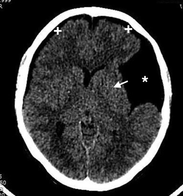
A 16-year-old female adolescent presented to the emergency department (ED) for a 3-week history of progressively worsening headaches associated with nausea and vomiting. She denied vision changes, photophobia, trauma, congestion, or fever. Steroid and antibiotic courses from an outside physician did not relieve symptoms. On examination in the ED, she had normal vital signs and a nonfocal neurological examination result. Computed tomography (CT) scan of the head was performed, revealing the diagnosis.
Diagnosis
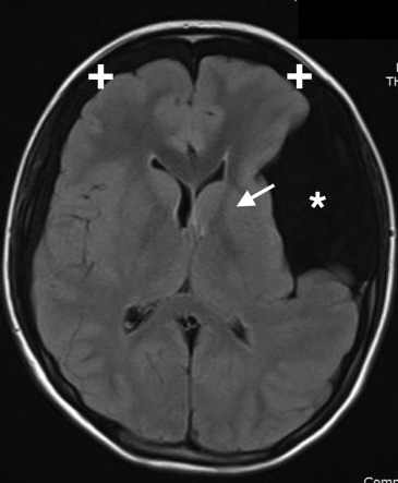
Ruptured arachnoid cyst. CT of the head revealed a large, ruptured, left-sided, frontotemporal arachnoid cyst with adjacent mass effect and bilateral subdural hygromas ( Figure 1 ). Magnetic resonance imaging (MRI) of the brain also confirmed the diagnosis ( Figure 2 ).


