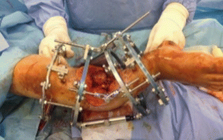
Fig. 16.1
Clinical picture demonstrates extensive tissue defect of the right lower limb after blast injury and primary debridement. Fracture stabilization using tubular external fixation frame
Repeated debridement procedure under general anesthesia was done 48 h later. Temporary acute shortening and anteromedial mal-angulation was performed to bring wound edges together and to close bone ends and the fracture site by viable soft tissues. Distal and proximal ends of the tibial bone were approximated by 8 cm shortening. The tibial bone fragments were angulated in a varus position 45°. Combination of acute limb mal-angulation with shortening allowed more complete coverage of the bone ends by the soft tissue (Fig. 16.2). During the operative procedure, peripheral pulse, capillary refill, and distal limb color were continually checked to ensure vascular viability.


Fig. 16.2
Clinical picture after performing acute shortening-angulation procedure. Conversion to hinged Ilizarov circular frame
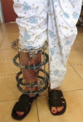
Local wound treatment was continued using method of negative pressure wound therapy (NPWT). Conversion to hinged Ilizarov circular frame was performed on day 10 using half pins of primary tubular external fixator. Skin grafting to granulating tissue was done 5 days later. After initial healing of the anteromedial wounds, gradual correction of the mal-angulation was begun. Over a period of 2 weeks, axial alignment was restored via isolated anteromedial distraction using the hinged circular Ilizarov external fixation frame, and full weight bearing to the injured limb with crutches was started (Fig. 16.3). Proximal tibial elongation corticotomy for distraction osteogenesis was done 1 week later. Graduate distraction osteogenesis in the Ilizarov frame by the rate ¼ mm × 4/d was started after 5 days. Control X-rays showed proper tibial alignment, and the girl was discharged to her home in Syria on day 70 with crutches and the Ilizarov device in situ.


