Chapter 42 Acute Renal Failure
1 How is acute renal failure (ARF) diagnosed?
ARF is a rapid loss of glomerular filtration rate (GFR) over a period of hours to a few days. This is usually determined by a sudden rise in plasma creatinine and blood urea nitrogen (BUN) levels. Until the patient achieves a steady state, the level of renal function cannot be assessed by the serum creatinine concentration. If a patient with previously normal renal function suddenly loses all renal function, serum creatinine will rise by only 1 to 2 mg/dL/day. However, patients with muscle wasting who make less creatinine may show smaller increases even with complete cessation of GFR. BUN is another indicator of decreasing glomerular filtration. Although a dramatic rise in BUN compared with creatinine may suggest a prerenal or obstructive (postrenal) cause, one must also consider the possibility that creatinine production by the patient is limited. Measurement of timed creatinine and urea excretion rates allowing for calculation of creatinine and urea clearance is sometimes indicated to clarify this point. In critically ill patients, decrease in urine output can be an indication of decreasing GFR. Increase in serum creatinine or decrease in urine output can be used to assess the risk, injury, and failure stages of ARF.
2 What features distinguish ARF from chronic renal failure?
When a patient is seen some time after the onset of ARF, this distinction may not be easy. Chronic renal failure is more likely than ARF to be associated with anemia, hypocalcemia, normal urine output, and small shrunken kidneys on ultrasound examination. A kidney biopsy may be warranted if the kidneys are of normal size. It has been reported that chronic, but not acute, renal failure may be associated with an increase in the serum osmolal gap (i.e., the difference between measured and calculated serum osmolality).
3 How is ARF classified?
The main categories are prerenal, intrarenal or parenchymal, and postrenal or obstructive (Table 42-1).
Table 42-1 Differential diagnosis of acute renal failure
Prerenal | Postrenal | Parenchymal |
---|---|---|
Dehydration | Ureter | Glomerular |
Impaired cardiac function | Bladder | Interstitial |
Vasodilation | Urethra | Allergic interstitial nephritis |
Renal vascular obstruction | Vascular | |
Hepatorenal syndrome | ATN |
ATN, Acute tubular necrosis.
4 How does examination of the urine help in the differential diagnosis of ARF?
Laboratory evaluation begins with careful examination of the urine. Concentrated urine points more to prerenal causes, whereas isotonic urine suggests parenchymal or obstructive causes. Typically, the urine sediment of patients with prerenal azotemia demonstrates occasional hyaline casts or finely granular casts. In contrast, the presence of renal tubular epithelial cells with muddy and granular casts strongly suggests acute tubular necrosis (ATN), microhematuria and red blood cell casts suggest glomerulonephritis, and white cell casts containing eosinophils suggest acute interstitial nephritis. Benign urine sediment is quite compatible with urinary obstruction.
5 What are the implications of urinary electrolytes in the differential diagnosis of ARF?
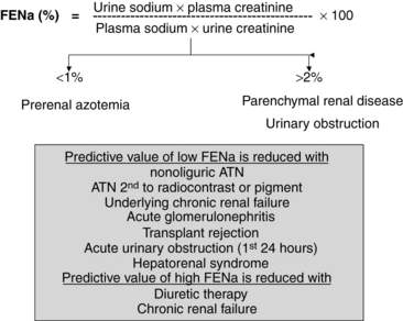
The determination of urine electrolyte and creatinine concentrations may be helpful in the differential diagnosis of ARF. When used with serum values, urinary diagnostic indexes can be generated. Understanding the concepts behind the interpretation of these indexes is easier and better than trying to remember specific numbers. Quite simply, if the tubule is working well in the setting of decreased GFR, tubular reabsorption of sodium and water is avid, and the relative clearance of sodium to creatinine is low. Conversely, if the tubule is injured and cannot reabsorb sodium well, the relative clearance of sodium to creatinine is not low. Therefore, with prerenal azotemia, the ratio of the clearance of sodium to the clearance of creatinine, which is also called the fractional excretion of sodium (FENa) (FENa = [Urinary sodium]/[Urinary creatinine] × [Plasma creatinine]/[Plasma sodium] × 100), is typically less than 1.0, whereas with parenchymal or obstructive causes of ARF, the FENa is generally greater than 2.0 (Fig. 42-1).

Figure 42-1 FENa, which measures the percentage of filtered sodium that is excreted in the urine, helps to differentiate between prerenal causes of renal failure and both parenchymal renal diseases (e.g., ATN, allergic interstitial nephritis) and urinary obstruction. Under specific conditions, the predictive value of a low FENa (to diagnose prerenal azotemia) or a high FENa (to exclude prerenal azotemia) is limited.
The FENa test is much less useful when patients do not have oliguria. In this setting, the specificity of a low FENa for prerenal azotemia is markedly diminished. In addition to nonoliguria, several causes of ATN, specifically dye-induced ATN or ATN associated with hemolysis or rhabdomyolysis, may typically be associated with a low FENa. Patients who have prerenal azotemia but have either persistent diuretic effect, chronic tubulointerstitial injury, or bicarbonaturia may have a relatively high FENa. In the last case, the fractional excretion of chloride, which is calculated in an analogous way, will be appropriately low (< 1%). Finally, the early stages of ARF from glomerulonephritis, transplant allograft rejection, or urinary obstruction may be associated with a low FENa.
6 What is the pathophysiology of ATN?
Renal ischemia, toxic injury to the kidney, or a combination of these insults can cause prolonged loss of renal function. Physiologically, decreased GFR must result from an alteration in glomerular hemodynamic factors, such as a decrease in the effective surface area or permeability of the glomerulus (Kf), a decrease in glomerular blood flow, or an abnormality in tubular integrity, including obstruction of tubular flow by cellular debris or back leak of ultrafiltrate through a porous tubule. In fact, each of these pathogenic features can be shown to be operant in some experimental models of ARF.
7 How does ATN evolve?
The major mechanism by which renal failure is induced may be different from the primary mechanism by which it is maintained. For example, in ischemic ARF, decreases in renal and glomerular blood flow may cause the initial loss of renal function. However, tubular necrosis, with its attendant obstructing debris and back leak of ultrafiltrate, maintains the low GFR. The tubular mechanisms are usually important in the maintenance of ARF from most causes seen clinically. Therefore pharmacologic efforts to improve renal blood flow are not, by themselves, generally effective in shortening the duration of ARF. Interestingly, modern ATN appears to recover much less quickly than when the syndrome was first described. This slow recovery may be related to repeated bouts of renal ischemia, which can be attributed to altered renal vasodilation related to the initial ATN insult. Therefore even mild degrees of hypotension should be avoided when treating patients with ATN.

Stay updated, free articles. Join our Telegram channel

Full access? Get Clinical Tree

