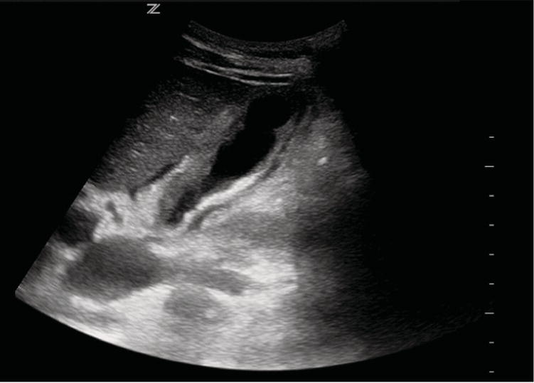
Marianna M. Fischmann1 and Fernanda Bellolio2 1 Department of Surgery, Pontifical Catholic University of Rio Grande do Sul, Porto Alegre, Brazil 2 Department of Emergency Medicine, Mayo Clinic, Rochester, MN, USA Acute cholecystitis is a common concern in patients presenting for the evaluation of abdominal pain, accounting for approximately 7% of patients presenting to the emergency department (ED) with abdominal pain.1 Patients typically present with pain localized to the right upper quadrant (RUQ), and the disease involves inflammation of the gallbladder, most commonly due to cystic duct obstruction by a gallstone. Gallstones themselves are relatively common, with a prevalence between 9% and 29% in United States patients, depending on ethnicity and gender.2 Risk factors for gallstones include age (>40 years), gender (women are more than twice as likely as men to have the disease), obesity (>120% of ideal body weight), pregnancy, and the use of oral contraceptives or estrogen replacement therapy. Figure 41.1 Acute cholecystitis with a thickened gallbladder wall and pericholecystic fluid seen on ultrasound. (Used with permission of Elke Platz, Heidi Kimberly & Dorothea Hempel, Department of Emergency Medicine, Brigham and Women’s Hospital.) Once present, gallstones can obstruct the cystic duct leading to the gallbladder inflammation that defines cholecystitis. Acute cholecystitis is typically diagnosed using ultrasound (with presence of gallstones, thickened gallbladder wall, pericholecystic fluid, and a sonographic Murphy sign; Figure 41.1), hepatobiliary cholescintigraphy (HIDA scan, with a lack of isotope accumulation in the gallbladder, indicating cystic duct obstruction), computed tomography (CT, with gallbladder wall thickening, pericholecystic fluid, and inflammation in the pericholecystic fat), or magnetic resonance imaging (MRI, with pericholecystic high signal intensity and visualization of the biliary tract).3 Secondary infection due to bile stasis commonly occurs, making empiric antibiotic therapy a mainstay of treatment.4 Definitive treatment requires cholecystectomy, and either percutaneous cholecystostomy5 or endoscopic gallbladder drainage via endoscopic retrograde cholangiopancreatography in patients thought to be too high risk for surgery. Acalculous cholecystitis, which occurs in up to 10% of cases of cholecystitis in adults and more than 50% of cases in children, can occur due to a number of infectious causes and has a higher morbidity and mortality.6 What is the accuracy of the history, physical examination, and laboratory testing for the diagnosis of acute cholecystitis? Trowbridge et al.7 conducted a review of studies comparing the physical exam, medical history, and lab tests in diagnosing acute cholecystitis. They included 17 studies that evaluated a total of 12 findings in the history and physical examination as well as five laboratory tests. They found that none of the tested findings, which included fever, Murphy sign, rebound tenderness, total bilirubin, liver function tests, and leukocytosis, had sufficient positive likelihood ratio (LR+) or negative likelihood ratio (LR−) to rule in or rule out the disease. Even RUQ tenderness, which had a summary LR− of 0.4, had a 95% confidence interval (CI) of 0.2–1.1. In a more recent systematic review, Jain et al.1 evaluated the diagnostic test accuracy of history, physical exam, and laboratory tests to predict if any of those elements would allow the diagnosis without utilizing imaging resources. When comparing point estimates, none of the characteristics including fever, RUQ mass, pain, tenderness or rebound, jaundice, Murphy’s sign, or vomiting had sufficiently low LR−’s to decrease the probability of cholecystitis. When evaluating laboratory findings, only one study met the inclusion criteria. The study analyzed the test characteristics of total bilirubin, showing that an elevated bilirubin increased the probability of the disease (LR+ = 5.8), but was not robust enough to significantly decrease the probability of cholecystitis (LR− = 0.64). The sensitivity and specificity of the test were 40% (95% CI 12–74%) and 93% (95% CI 77–99%), respectively. The authors suggested that the development of a clinical decision rule with multiple parameters (not including imaging) could be useful for the correct diagnosis, since no single predictor of history, physical examination, or laboratory test is sufficient to diagnose or rule out acute cholecystitis. The last update of the Tokyo Guideline for the management of acute cholecystitis (TG18) utilized an expert consensus methodology to integrate the diagnostic modalities.8 They suggest that if at least one of the local signs of inflammation (Murphy’s sign, RUQ mass/pain/tenderness) and one systemic sign of inflammation (fever, elevated C‐reactive protein, elevated white blood cell count) are present, the patient should be classified as suspected cholecystitis. However, a confirming imaging (ultrasound, HIDA, CT, or MRI) is necessary to achieve definite diagnosis. Validation studies found that the diagnostic accuracy from this diagnostic criterion ranges from 60% to 94%. What is the accuracy of CT, MRI, ultrasound, or nuclear medicine scans for the diagnosis of acute cholecystitis? Ultrasound is the first imaging option for patients with suspected cholecystitis.3 HIDA was considered the criterion standard for diagnosing acute cholecystitis for a long time. In a single‐center retrospective analysis of patients suspected of having acute cholecystitis (who had both ultrasound and HIDA scan tests ordered simultaneously), the performance characteristics of the two tests were compared.9 It was standard practice at the study hospital for both studies to be ordered together when the diagnosis was considered. Consecutive patients were included, and patients were excluded if a test was ordered based on a prior test to minimize bias based on the initial tests’ results. The final diagnosis was determined using surgical, pathology, autopsy reports, or a clinical diagnosis for those who did not undergo surgery. A total of 107 patients were examined, 32 (30%) of whom had a final diagnosis of acute cholecystitis. Using data provided in their study, the performance characteristics of the two tests are shown in Table 41.1. The authors concluded that HIDA was superior to ultrasound in diagnosing acute cholecystitis. They suggest that, since the costs of each of these studies at the time in their institution were similar, the decision should be based on availability and diagnostic performance. A systematic review and meta‐analysis were performed by Kiewiet et al.10 to obtain summary estimates of diagnostic accuracy for different imaging modalities (Table 41.2). Data regarding CT accuracy could not be summarized because only one study met the inclusion criteria. In that study, CT had a sensitivity of 94% (95% CI 73–99%) and a specificity of 59% (95% CI 42–74%) for the detection of acute cholecystitis. For MRI, the summary estimate of sensitivity was 85% (95% CI 66–95%) and specificity was 81% (95% CI 69–90%). Ultrasound had a sensitivity and specificity of 81% (95% CI 75–87%) and 83% (95% CI 74–89%), respectively. HIDA had a sensitivity of 96% (95% CI 94–97%) and specificity of 90% (95% CI 86–93%). The summarized estimates were significantly higher for HIDA than for ultrasound and the authors suggested that HIDA cholescintigraphy has the highest diagnostic accuracy of all imaging modalities. Because of its lower availability, and associated radiation and contrast dye use, HIDA should only be considered in clinically equivocal cases.11,12 Table 41.1 Test characteristics of hepatobiliary scintigraphy (HIDA) scan and ultrasound for cholecystitis Source: Data from [9].
Chapter 41
Acute Cholecystitis
Background
Clinical question
Clinical question
Acute biliary disease (+)
Acute biliary disease (−)
Totals
HIDA (+)
28
5
33
HIDA (−)
Related posts:
![]()
Stay updated, free articles. Join our Telegram channel

Full access? Get Clinical Tree

 Get Clinical Tree app for offline access


