ACUTE AORTIC PATHOLOGY
CASE SCENARIO
A 65-year-old male with a past medical history notable for hypertension, hyperlipidemia, and a 40 pack-year smoking history complicated by chronic obstructive pulmonary disease (COPD) presents to the emergency room with 2 hours of sudden-onset abdominal and lower back pain. Shortly after the pain began, he felt dizzy and nauseated. He was subsequently brought to the emergency room by ambulance.
Upon evaluation, the patient is found to be tachycardic and hypotensive with a systolic blood pressure of 90. He is slightly somnolent but oriented to person, place, and time. His abdomen is distended and mildly tender to palpation without overt signs of peritonitis. A prominent pulsatile midline mass is also noted. The bilateral flanks demonstrate ecchymosis. Femoral, popliteal, and distal lower extremity pulses are diminished but palpable, and all extremities are mottled and cool. His labs are notable for a hematocrit of 33, white blood cell count of 12, BUN of 20, and Cr of 1.4.
EPIDEMIOLOGY
Acute diseases of the aorta present life-threatening emergencies. The following discussion aims to equip the reader with the understanding required for prompt diagnosis and initial management of aortic aneurysm and dissection.
 Abdominal Aortic Aneurysm (AAA)
An arterial aneurysm is a localized, permanent, focal dilation that exceeds the normal vessel diameter by 50% or greater.1,2 Measurements taken with a variety of radiographic and in situ techniques have demonstrated that the normal caliber of the abdominal aorta varies in size by location and by gender. The supraceliac aorta is largest, with a diameter ranging from 2.50 to 2.72 cm in men and 2.10 to 2.31 cm in women, whereas the infrarenal aorta ranges from 1.99 to 2.39 cm in men and 1.66 to 2.16 cm in women.3 In general, however, an abdominal aortic aneurysm is defined by an aortic diameter greater than or equal to 3 cm, or a 1.5-fold increase in a patient’s normal aortic diameter.4 In the vast majority of cases, the isolated area of aneurysmal dilation is confined to the infrarenal aorta (~80%).4,5
The incidence of AAA ranges from approximately 1.5% to just over 3% in various studies,4,5 and the rate of aneurysm formation is two- to six-fold higher in men compared to women.3 Rates as high as 10% to 15% have been noted in populations with a strong family history of aneurysmal disease and in long-term smokers.6
 Aortic Dissection
The incidence of acute aortic dissection is approximately 3 per 100,000 person-years.6,7 Dissections typically occur between the sixth and eighth decades of life and affect males more commonly than females. Accurate diagnosis and timely intervention are paramount to minimize morbidity and mortality. Studies have demonstrated that without treatment, most patients with acute dissection die within 3 months of diagnosis.7 Mortality rates for acute dissection worsen with time, with a rate of 22.7% in the first 6 hours, 50% within 24 hours, and 68% within the first week.7 Those who survive the early phase often develop aneurysmal degeneration of the dissected aorta, resulting in high rates of rupture by 5 years.7
PATHOPHYSIOLOGY
 Abdominal Aortic Aneurysm
The underlying etiology of AAA formation is not clearly understood, but is likely multifactorial. At a cellular level, atherosclerosis has traditionally been invoked as a driving force in aneurysm formation; however, more recent studies demonstrate significant contributions from inflammation, as well as enzymatic breakdown of the arterial wall through matrix metalloproteinases, collagenases, and other proteases.8 Genetics have also been implicated, particularly in the case of connective tissue disorders. Finally, hemodynamic perturbations such as hypertension may contribute to aneurysm formation, while distal iliac atherosclerotic disease may increase resistance to outflow and elevate pressures within the aneurysm sac.
Mycotic aneurysms are often a misnomer, and a misunderstood entity. While the association between vascular infection and aneurysms is well documented, at least three separate diagnostic entities should be acknowledged: true mycotic aneurysms, superimposed bacterial infection of an existing aneurysm, and bacterial arteritis leading to aneurysm.
True mycotic aneurysms are a consequence of septic emboli. An embolus becomes lodged within small vessels, damaging the vessel wall. In larger vessels such as the aorta, septic emboli may lodge in the vasovasorum, resulting in ischemia of the aortic wall and bacterial colonization. In both cases, the most common source of septic emboli is infectious endocarditis. Organisms associated with this process include Streptococcal and Staphylococcal species; however, numerous others have been reported in the literature.
Unlike mycotic aneurysms, which develop as a result of septic emboli, existing aneurysms can themselves become infected during episodes of bacteremia, when bacteria invade the damaged intimal wall. Similarly, bacterial invasion of the inflamed intima in microbial arteritis can lead to aneurysm formation. Perhaps the most important organism noted in cases of bacterial aortitis with aneurysm formation is Salmonella species. This is thought to result from Salmonella’s ability to invade the aortic endothelium in areas with atherosclerotic burden, although intimal invasion in areas without disease is also possible.9
Similar to aneurysm formation, the exact pathophysiology of rupture is poorly understood; however, autopsy studies have demonstrated that approximately one third of aneurysms ultimately progress to rupture.10 Rupture occurs when pressure exerted on the inner surface of the aortic wall exceeds the capacity of the vessel to withstand such force. This breaking point is achieved at a much lower pressure in an aorta with aneurysmal degeneration, and the likelihood of rupture increases as the aneurysm grows. This relationship is grossly estimated by the law of LaPlace, which states that the wall tension of the aneurysm sac is proportional to the radius of the sac multiplied by the transmural pressure, and inversely proportional to wall thickness. In the case of mycotic and infected aneurysms, compromised wall integrity translates into rupture at smaller sizes and lower pressures.
 Aortic Dissection
Similar to AAA, the exact etiology of aortic dissection is poorly understood, although hypertension contributes significantly. Cystic medial necrosis resulting in degeneration of the aortic media has also been implicated. Connective tissue disorders such as Marfan’s and Ehlers-Danlos syndromes, as well as the aortopathy associated with bicuspid aortic valves, result in higher rates of dissection. As opposed to AAA, atherosclerosis does not appear to play a major role. In fact, the inflammation associated with the deposition of material between the intima and media in atherosclerosis results in a fusion of these two layers, which may actually arrest progression of the dissection.7
A dissection occurs when an area of decreased intimal integrity allows blood to penetrate the intima and dissect through the potential space between the intima and media. The result is formation of a false aortic lumen, contained medially by the outer side of the intima and laterally by the media and adventitia. Because the false lumen has thinner walls but is initially exposed to the same mean arterial pressure as the true lumen, the law of LaPlace dictates that the false lumen should expand while the true lumen collapses, such that the wall tension of both lumens equilibrates. Collapse of the true lumen may in turn impair end-organ perfusion. Additionally, the distal aspect of the dissection flap, or thrombus formation within the false lumen, may obstruct the ostium of an arterial branch, with reduced blood flow and resultant ischemia, often termed a malperfusion syndrome.
Two major classification systems—Stanford and DeBakey—describe the origin and extent of an aortic dissection. The Stanford classification system is more commonly used, and describes the dissection in relation to involvement of the ascending aorta. Type A dissections originate in the ascending aorta, although they can also involve the descending, whereas type B dissections are limited to the descending aorta.
The DeBakey classification system has three categories: Types I, II, and III. Type I dissections originate in the ascending aorta and extend into both the ascending and descending aorta. Type II dissections, in contrast, originate and remain isolated within the ascending, and Type III occur within the descending aorta exclusively. DeBakey Type I and II are both grouped under Stanford A, and Debakey Type III is the equivalent of a Stanford B (Table 20–1 and Figure 20–1).

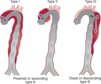
Figure 20–1 Illustration of the Stanford and Debakey classifications of aortic dissection. (Reproduced with permission from Doherty GM Current Diagnosis and Treatment: Surgery, 13th Ed. New York, NY: McGraw-Hill;2010.)
Unlike aneurysms, in which rupture is the main source of morbidity and mortality, dissections wreak havoc by compromising end-organ perfusion. Involvement of the visceral segment in a dissection flap can instigate life-threatening mesenteric ischemia requiring emergent surgical exploration. Additionally, type A dissections can disrupt the aortic annulus, prompting acute aortic regurgitation and cardiogenic shock. In some cases, a type A dissection can extend into the pericardium, with resultant hemopericardium, tamponade, and death. These devastating complications are associated with high mortality, and as such the vast majority of type A dissections require emergent surgical intervention.
Although dissections are less likely to rupture than aneurysms, most chronic type B dissections that are managed medically will degenerate into thoracoabdominal aneurysms as a result of ongoing false lumen expansion over time, and will ultimately progress to rupture if not resected.
CLINICAL PRESENTATION
 Abdominal Aortic Aneurysm
The clinical presentation of abdominal aortic aneurysms ranges from asymptomatic, to shock with multisystem organ dysfunction, to free rupture with sudden death. The following section reviews the presentation of asymptomatic and symptomatic aneurysms, aneurysms with contained and free rupture, and mycotic aneurysms.
Astute clinicians who notice a prominent abdominal aortic pulsation during routine physical examination occasionally discover asymptomatic aneurysms. More often, though, an asymptomatic aneurysm is identified on a screening ultrasound, or incidentally picked up on a computed tomography (CT) scan of the abdomen performed for workup of abdominal pain.
Symptomatic aneurysms can present with vague abdominal pain related to mass effect from the growing aneurysm, pain from rapid expansion, thrombosis of the abdominal aorta, or embolic sequelae. In the case of mass effect, patients describe symptoms related to compression of a specific organ, such as early satiety from gastric or bowel compression, hydronephrosis from ureteral obstruction, or lower extremity edema from vena cava or iliac vein compression. Rapid expansion may present with vague to severe abdominal pain, while severe tearing pain that radiates to the back is classic for dissection. Thrombotic and embolic complications may develop as mural thrombus accumulates within the aneurysm sac and embolizes to the lower extremities, manifesting as acute limb-threatening or digital ischemia (as in the case of “blue toe syndrome”). Complete thrombosis of the abdominal aorta is a true surgical emergency, and can result in devastating visceral and bilateral lower extremity ischemia.
Contained rupture occurs when blood breaches the aortic wall, but surrounding tissues tamponade the hemorrhage. In most cases, this translates into rupture into the retroperitoneum. Patients with contained rupture often present with severe abdominal and or flank pain, with or without accompanying hypotension and end-organ malperfusion.
Free rupture occurs when hemorrhage is not tamponaded by surrounding structures, and has an estimated mortality rate of 90% secondary to exsanguination.11 This often results from rupture into the peritoneal cavity; however, rupture into adjacent visceral structures presenting as massive gastrointestinal bleeding, or into venous structures resulting in acute congestive heart failure in the case of an aorto-caval fistula, can also be seen.11
Mycotic aneurysms have subtle clinical findings. Patients may complain of systemic symptoms of low-grade infection such as fatigue, malaise, and recurrent fevers or chills. The aneurysm itself may be quite tender to palpation on physical examination, even if intact. In the case of true mycotic aneurysms in the setting of infective endocarditis, physical examination findings related to the latter, including new murmurs, splinter hemorrhages, Janeway lesions, and Osler’s nodes, may be the predominant findings. In some cases, patients may present with overt septic physiology.
 Aortic Dissection
Dissections of the thoracic aorta present with chest and back pain that is described as tearing or ripping in quality. In general, additional clinical symptoms, examination findings, and laboratory abnormalities are related to mass effect from the expanding false lumen or to ischemia of an organ, as described above. In the case of the former, a Horner’s syndrome or a hoarse voice may develop from compression of the sympathetic trunk or the recurrent laryngeal nerve, respectively. Malperfusion syndromes, meanwhile, can affect any vascular supply arising directly from the aorta. Proximally, dissections can extend into the coronary arteries or occlude their ostia, resulting in myocardial ischemia that may present with chest pain superimposed on pain from the dissection, or electrocardiogram (EKG) changes with elevated cardiac biomarkers. Arch involvement may cause unequal blood pressures in the upper extremities and/or neurologic deficits related to decreased cerebral blood flow. Malperfusion of intercostal branches of the descending thoracic aorta may result in neurologic deficit from spinal cord ischemia, and involvement of the abdominal aorta may present with a clinical picture of bowel ischemia or acute renal failure. If the dissection extends beyond the iliac bifurcation, unequal femoral and lower extremity pulses may be appreciated. Finally, as mentioned earlier, dissections involving the proximal ascending aorta or dissections of the distal arch that progress in a retrograde fashion may compromise the structural integrity of the aortic valve annulus, resulting in acute aortic insufficiency and its sequelae such as cardiogenic shock and pulmonary edema. The dissection may also extend into the pericardial space, where hemopericardium and tamponade may ensue.
DIFFERENTIAL DIAGNOSIS
 Abdominal Aortic Aneurysm
Because of the retroperitoneal location of the abdominal aorta, pain related to an unruptured symptomatic aneurysm, or pain resulting from contained rupture is often vague and unlikely to present with the somatic pain associated with peritoneal irritation. The differential is therefore extremely broad. Accurate diagnosis relies heavily on consideration of the patient’s history, including known cardiovascular risk factors, and further evaluation with diagnostic studies (Table 20–2).
 Aortic Dissection
The differential diagnosis for aortic dissections should be thought of in two ways: a clinical differential diagnosis, and a radiographic differential diagnosis. Clinically, myocardial infarction and pulmonary embolism may present very similarly to dissection, and this condition must be ruled out as part of the process. Any patient arriving to the emergency department with risk factors for either coronary disease or dissection should undergo cardiac biomarker testing and evaluation with an EKG at the very minimum. It should be noted, however, that myocardial infarction can be secondary to a dissection, and therefore further imaging should be considered if there is ongoing high suspicion for dissection. Less common conditions that may mimic dissection include musculoskeletal pain or esophageal pathology such as spasm; however, these are clearly diagnoses of exclusion.
The radiographic differential diagnosis for aortic dissection includes mural thrombus and penetrating aortic ulcers. In the case of the former, mural thrombus in a normal-caliber aorta or thrombus within an aneurysm may give the impression of a thrombosed false lumen in association with a dissection. Penetrating aortic ulcers may also mimic a local dissection as contrast penetrates through the aortic intima (Table 20–3).

WORKUP AND CHOICE OF IMAGING
 Abdominal Aortic Aneurysm
The most noninvasive and least expensive imaging modality for identification of AAA is B-mode ultrasound, with a sensitivity near 100%. In fact, because of the relative ease of acquisition and excellent accuracy that ultrasound offers, and the significant morbidity and mortality that can be avoided with elective AAA repair, routine screening is now recommended for patients with significant risk factors, the most important of which are male gender, age greater than 65, and a history of smoking.11,12
Ultrasound, however, is limited by operator dependence. Visualization of the suprarenal aorta can prove challenging, while the infrarenal aorta can be obscured easily by overlying bowel gas. Moreover, the ability to distinguish between ruptured and unruptured aneurysms is quite difficult in the absence of obvious intraperitoneal fluid. Therefore, in order to accurately diagnose aneurysm rupture, and for operative planning, imaging with computed tomographic angiography (CTA) or conventional angiography is required.
Although conventional angiography was previously defined as the gold standard for diagnosis, the speed and accuracy of modern helical CT scanners have made CTA the current standard. Images provide more accurate visualization of aneurysm morphology and measurements when compared to B-mode ultrasound.
In the modern era of endovascular management, preoperative CTA is essential for operative planning. In addition to the above-mentioned advantages, CTA can accurately differentiate a ruptured from a, unruptured aneurysm by visualizing. Finally, with regard to differential diagnosis and other intra-abdominal pathology that may mimic symptoms of ruptured or unruptured AAA, CTA has the advantage of visualizing other processes throughout the abdominal cavity.
Obtaining arterial phase images does not detract from the standard abdominal CT scan that is often routinely ordered in emergency departments to assist with evaluation of abdominal pain, as long as the usual delayed phase images are also obtained following the arterial one. It should be noted, however, that if there is any concern for AAA or other intra-abdominal vascular pathology, a CTA should be protocoled at the time of the initial scan. Acquiring arterial phase images after a “routine” abdominal requires an additional contrast load, creating an unacceptable risk of contrast-induced nephropathy.
Similar to noninfected aneurysms, mycotic aneurysms are easily detected by ultrasonagraphy; however, this imaging modality cannot identify underlying infection. Likewise, CTA will clearly demonstrate the aneurysm, but cannot reliably indicate whether the aneurysm is mycotic, although aneurysm morphology and secondary signs of infection can be suggestive.13
Magnetic resonance imaging (MRI) has been reported in the literature as a potential modality to evaluate for mycotic aneurysms, as certain sequences may demonstrate inflammation. However, as with CTA, detection is often dependent on evidence of nearby tissue inflammation, for example, vertebral body enhancement.14 At our institution, we often employ tagged white blood cell scans in order to look for aneurysmal enhancement suggestive of superimposed infection; however, these studies are often more sensitive in the setting of infected synthetic graft material rather than native aorta.
 Aortic Dissection
Patients arriving in the emergency department with a chief complaint of chest pain will inevitably undergo a chest x-ray (CXR), which may demonstrate a widened mediastinum associated with dissection, though this finding alone is neither sensitive nor specific. Portable antero-posterior (AP) CXRs have a tendency to widen the mediastinal and cardiac silhouettes, and obese patients may have a widened mediastinum without concomitant aortic pathology. Accurate diagnosis therefore depends on more definitive imaging.
As with the diagnosis of AAA, CTA has become the standard for the diagnosis of dissections. In fact, most institutions offer a “dissection protocol” CT with arterial phase images of both the thoracic and abdominal aorta. CTA may offer improved sensitivity over traditional aortography, which can occasionally fail to visualize a thrombosed false lumen.
In addition to confirming the presence of a dissection, CTA can further elucidate the extent of the dissection and involvement of arterial branches, and differentiate if arterial branches arise from the true or false lumen. In some cases, CTA can demonstrate evidence of end-organ malperfusion, manifesting as wall thickening and pneumatosis in the case of the bowel, or hypoattenuation in the case of an ischemic or infarcted kidney. Liver or spleen ischemia may be seen as heterogeneity, with some areas of the organ “lighting up” less than others. Finally, when dissection extends into the pericardium, hemopericardium and occasionally tamponade (concavity of the right atrium or right ventricle) can be seen on CTA.
CTA exposes the patient to radiation and the risks associated with intravenous contrast, namely, contrast-induced nephropathy or anaphylaxis. When end-stage renal disease or contrast allergy precludes the use of CTA, other modalities such as transesophageal echocardiography (TEE) and MRI can be employed. TEE has a sensitivity for dissection ranging from 35% to 80% and specificity ranging from 40% to 95%.7 Like other ultrasound-based diagnostic studies, the sensitivity and specificity of TEE is operator dependent. The benefit of TEE in skilled hands, however, is rapidity of image acquisition.
For patients with mild to moderate renal insufficiency or intravenous contrast allergy, MRI is an excellent alternative for diagnosis, with sensitivity and specificity rates approaching 100%. Limitations include much longer examination times when compared with CTA, and decreased availability in some institutions. Gadolinium use is also limited in patients with significant renal insufficiency.
IMAGING FINDINGS
 Abdominal Aortic Aneurysm
Ultrasound
As mentioned previously, ultrasound cannot accurately determine the presence of rupture, particularly when rupture is contained to the retroperitoneum. In cases where there is free intra-peritoneal rupture, ultrasound may demonstrate free fluid in the hepatorenal and splenorenal fossae similar to a positive focused assessment with sonography for trauma (FAST) examination. Please refer to Chapter 3, “Bedside Ultrasound for Surgeons” for a complete discussion and sample images.
CTA
AAA is recognizable on CTA as a dilation of the aorta >3 cm. There may be significant associated mural thrombus and calcifications, and the aorta may adopt a tortuous appearance (Table 20–4 and Figures 20–2 and 20–3).


