Abdominal Trauma
Ulises Torres
I. OVERVIEW
A. Background.
1. The abdomen is frequently injured after both blunt and penetrating trauma.
2. Abdominal trauma occurs in 20% of civilian injuries requiring surgery.
3. Half of all preventable deaths are related to suboptimal management of abdominal trauma.
4. Physical examination is often inadequate to identify or exclude intraabdominal injuries (especially blunt trauma).
5. Multiple factors, including mechanism of injury, body region injured, hemodynamic and neurologic status, and associated injuries, influence the diagnostic approaches used and outcome of abdominal injuries.
B. Etiology.
1. Blunt trauma (the most common etiology) is caused by motor vehicle crashes (MVCs), motorcycle crashes, falls, and assaults and to pedestrians struck by vehicles.
2. Penetrating abdominal trauma is usually caused by gunshot or stab wounds.
3. Almost all causes are preventable.
C. Diagnosis.
1. History and physical examination.
a. Trauma history: Allergies, Medications, Past medical/surgical history, Last meal, Events surrounding the injury (AMPLE).
b. Knowing the mechanism of the trauma is imperative in order to suspect the different intra-abdominal injuries.
c. Suggestive clinical findings include abdominal wall contusions (seat-belt sign), pain, tenderness, or unexplained hypotension (Fig. 113-1).
d. Peritoneal signs are absent in 40% of patients with significant abdominal injuries.
e. Rectal examination for gross blood, bony fractures, tone, or urethral injury, although it is not a sensitive test.
f. Foley catheterization is appropriate when no urethral injury is suspected: hematuria indicates injury to the bladder, ureters, or kidneys; in those cases, retrograde urethrogram is indicated.
g. Physical findings are unreliable for patients with an abnormal sensorium secondary to head trauma, spinal cord injury, intoxication, or distracting pain from another source.
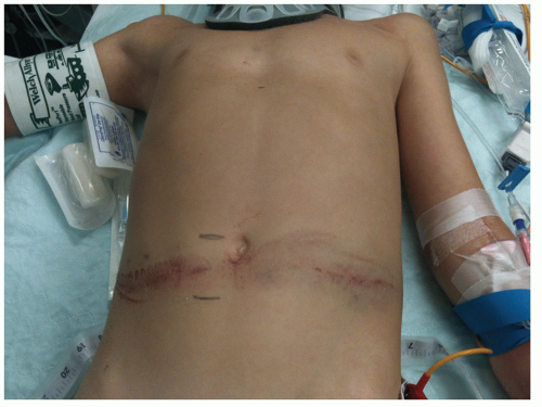 Figure 113-1. The seat belt sign, clearly demonstrated above, should trigger further investigation and observation. |
2. Plain radiographs.
a. Chest x-ray may reveal pneumoperitoneum; a gastric bubble or bowel in the chest is consistent with a ruptured diaphragm, and lower rib fractures suggest underlying liver or spleen injury.
b. Abdominal x-ray has no role in the assessment of abdominal trauma with the exception of defining the trajectory of a projectile in order to choose the incision by the surgery team.
3. Computed tomography (CT).
a. Abdomen and pelvic CT is the mainstay of diagnosis for abdominal and retroperitoneal injury in the hemodynamically stable patient.
b. With intravenous contrast material, CT can detect arterial contrast extravasation.
c. Oral contrast is not necessary for trauma CT; does not increase sensitivity.
d. Sensitivity rates are between 92% and 97.6% and specificity rates as high as 98.7%. Negative predictive value up to 99%.
e. Unreliable in identifying hollow visceral injuries.
f. Contraindicated in hemodynamically unstable patients (who would benefit from intervention, rather than diagnostic testing).
4. Focused assessment with sonography for trauma (FAST).
a. Includes four areas of examination:
i. Right upper quadrant: Morrison pouch between the right kidney and liver.
ii. Left upper quadrant: between the left kidney and spleen.
iii. Subxiphoid: pericardium (alternatively transsternal).
iv. Suprapubic: perivesicular spaces.
5. FAST has mostly replaced diagnostic peritoneal lavage (DPL) and gained acceptance in the evaluation of abdominal trauma, especially in the hemodynamically unstable patient; primarily used to detect occult intra-abdominal blood. DPL and/or FAST usage depends on the skill and experience of the clinician.
a. FAST is accurate in detecting intra-abdominal blood (94% to 96%) in experienced hands.
b. Advantages include being rapid, portable, and noninvasive; disadvantages include low specificity for source of hemorrhage and high operator dependence.
c. A normal FAST does not exclude injuries of hollow viscus and retroperitoneal injuries.
d. With appropriate training, FAST can be effectively performed by nonradiologists.
e. The FAST examination can be easily used at the bedside at any time after the initial trauma.
6. Laparoscopy.
a. Use of diagnostic laparoscopy in blunt abdominal trauma is a developing field.
b. Difficult to adequately examine small bowel and retroperitoneum.
c. Accurate in determining peritoneal or diaphragmatic penetration from stab wounds or tangential gunshot wounds.
7. Nonoperative management (NOM) and serial examinations.
a. Can be used for some blunt trauma and superficial stab wound patients with normal mental status and hemodynamic stability.
D. Treatment.
1. For blunt abdominal trauma, treatment is dictated by the specific injuries present.
2. Operative exploration is required in all patients with anterior abdominal gunshot wounds because visceral injury is present in >90% of cases.
3. For stab wounds, exploration is necessary in the presence of peritoneal signs, hemodynamic instability, evisceration of abdominal contents, or confirmed peritoneal penetration.
4. Unexplained hypotension with physical signs of abdominal trauma may warrant exploration.
II. SPECIFIC INJURIES
A. Liver and porta hepatis.
1. Background.
a. The liver is the most commonly injured organ in blunt abdominal trauma.
b. Spontaneous hemostasis is observed in >50% of small hepatic lacerations at the time of laparotomy.
c. Most liver injuries heal without intervention.
d. Overall mortality rate ranges from 8% to 10% depending on the number of associated injuries and injury severity.
e. Injuries are graded from I (minor) to V (severe), ranging from minor tears to major lobar disruptions and avulsion from the inferior vena cava.


