A 65-year-old woman presented to the emergency department with worsening dyspnea on exertion. She had a history of deep venous thrombosis and within the past month had been traveling extensively. Examination was notable for dyspnea without unilateral leg swelling or jugular venous distention, a room air oxygen saturation of 92%, and tachycardia of 102 beats/min. D-dimer level was elevated, at 2,636 ng/mL. Computed tomography (CT) pulmonary angiography confirmed the diagnosis.
Diagnosis
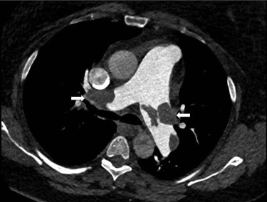
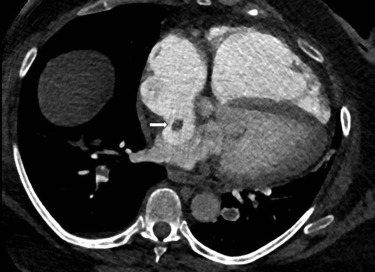
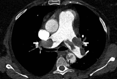
Acute submassive pulmonary embolism with systemic embolization through a patent foramen ovale. CT pulmonary angiography demonstrated bilateral pulmonary embolism ( Figure 1 ) and thrombus extending into the left atrium through a patent foramen ovale ( Figure 2 ). On the first day of hospital admission, a CT angiogram of the chest, abdomen, and pelvis showed aortic embolization ( Figure 3 ). She received anticoagulation and underwent surgical embolectomy and patent foramen ovale closure.
Related posts:
 Adolescent Male With Right Shoulder Pain After Football Injury
 Association Between the Opening of Retail Clinics and Low-Acuity Emergency Department Visits
Bending the Curve of Health Trajectories for Older Adults Discharged From the Emergency Department
Yet Another
Sedating the Agitated Patient: A Moving Target?
Classified 2017 Advertising Rates & Information
Association Between the Opening of Retail Clinics and Low-Acuity Emergency Department Visits
Bending the Curve of Health Trajectories for Older Adults Discharged From the Emergency Department
Yet Another
Sedating the Agitated Patient: A Moving Target?
Classified 2017 Advertising Rates & Information
Stay updated, free articles. Join our Telegram channel

Full access? Get Clinical Tree


