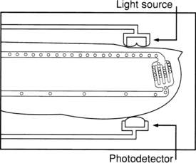
PROCEDURE 17 • Oxygen saturation is an indicator of the percentage of hemoglobin saturated with oxygen at the time of the measurement. The reading, obtained with standard pulse oximetry, uses a light sensor that contains two sources of light (red and infrared) absorbed by hemoglobin and transmitted through tissues to a photodetector. The infrared light is absorbed by the oxyhemoglobin, and the red light is absorbed by the reduced hemoglobin. The amount and type of light transmitted through the tissue is converted to a digital value that represents the percentage of hemoglobin saturated with oxygen (Fig. 17-1). • Oxygen saturation values obtained with pulse oximetry (SpO2) represent one part of a complete assessment of a patient’s oxygenation status and are not a substitute for measurement of arterial saturation of oxygen (SaO2) or of ventilation (as measured with arterial partial pressure of carbon dioxide [PaCO2]). • The accuracy of SpO2, measurements requires consideration of many physiologic variables. Patient variables include the following: • A complete assessment of oxygenation includes evaluation of oxygen content and delivery, which includes the following parameters: arterial partial pressure of oxygen (PaO2), SaO2 hemoglobin, cardiac output, and, when available, mixed venous oxygen saturation. • Normal oxygen saturation values are approximately 97% to 99% in a healthy individual breathing room air. An oxygen saturation value of 95% is clinically accepted in a patient with a normal hemoglobin level. With a normal blood pH and body temperature, an oxygen saturation value of 90% is generally equated with a PaO2 of 60 mm Hg. • Tissue oxygenation is not reflected by arterial or oxygen saturation obtained with pulse oximetry. • The affinity of hemoglobin with oxygen may impair or enhance oxygen release at the tissue level. • Oxygen saturation values may vary with the amount of oxygen usage or uptake by the tissues. In some patients, a difference is seen in SpO2 values at rest compared with values during activity, such as ambulation or positioning. • Oxygen saturation does not directly reflect the patient’s ability to ventilate. The true measure of ventilation is determination of the PaCO2 in arterial blood. Use of SpO2 in a patient with obstructive pulmonary disease may result in erroneous clinical assessments of condition. As the degree of lung disease increases, the patient’s drive to breathe may shift from an increased carbon dioxide stimulus to a hypoxic stimulus. Enhancing the patient’s oxygenation and increasing the SpO2 may limit the ability to ventilate. The normal baseline SpO2 for a patient with known severe restrictive disease and more definitive methods of determination of the effectiveness of ventilation must be assessed before consideration of interventions that enhance oxygenation. • Any discoloration of the nail bed or obstruction of the nail bed can potentially affect the transmission of light through the digit. The impact of dark nail polish, such as blue, green, brown, or black colors,4,5,11,17 has been reported to limit the transmission of light and thus impact the SpO2, although a recent study showed that fingernail polish does not cause a clinically significant change in the pulse oximeter readings in healthy individuals.15 If the nail polish cannot be removed and is believed to be affecting the accuracy of the reading, the sensor can be placed in a lateral side-to-side position on the finger to obtain readings if no other method of sampling the arterial bed is available.5,15 Bruising under the nail can limit the transmission of light and result in an artificially decreased SpO2 value. Pulse oximetry has not been shown to be affected by the presence of an elevated bilirubin.2 The presence of acrylic fingernails may impair the accuracy of the pulse oximetry reading, and removal of the nail covering may be necessary to ensure accurate measurement,9 although unpolished acrylic nails have been proven not to affect pulse oximetry readings.14
Oxygen Saturation Monitoring with Pulse Oximetry
PREREQUISITE NURSING KNOWLEDGE
 Presence of dyshemoglobinemias (i.e., carboxyhemoglobinemia after carbon monoxide exposure)
 Arterial blood flow to the vascular bed
 Temperature of the digit or the area where the oximetry sensor is located
 Fraction of inspired oxygen (percentage of inspired oxygen)
 Evidence of ventilation-perfusion mismatch
 Amount of ambient light seen with the sensor
 Oxygen is more readily released to the tissues when pH is decreased (acidosis), body temperature is increased, PaCO2, is increased, and 2,3-diphosphoglycerate levels (a by-product of glucose metabolism that facilitates the dissociation of oxygen from the hemoglobin molecule to tissue) are increased (decreased oxygen affinity).
 When hemoglobin has greater affinity for oxygen, less is available to the tissues (increased oxygen affinity). Conditions such as increased pH (alkalosis), decreased temperature, decreased PaCO2, and decreased 2,3-diphosphoglycerate (as found in stored blood products) increase oxygen binding to the hemoglobin and limit its release to the tissue.
![]()
Stay updated, free articles. Join our Telegram channel

Full access? Get Clinical Tree


17: Oxygen Saturation Monitoring with Pulse Oximetry


