Fig. 16.1
FloTrac monitor showing four different parameters: cardiac output (CO), cardiac index (CI), stroke volume (SV), and stroke volume variation (SVV)
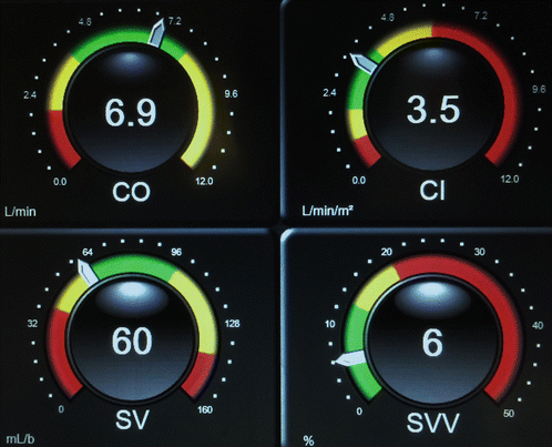
Fig. 16.2
FloTrac monitor showing four different parameters: cardiac output (CO), cardiac index (CI), stroke volume (SV), and stroke volume variation (SVV)
- 1.
What do the figures show?
- 2.
What is the importance of monitoring cardiac output?
- 3.
What are the advantages of the FloTrac/EV1000 system?
- 4.
How does the FloTrac estimate stroke volume?
- 5.
What are the limitations of the FloTrac?
- 6.
What other minimally invasive monitors are available?
- 7.
What is stroke volume variation?
- 8.
Why is the stroke volume higher during the inspiratory phase of the respiratory cycle?
- 9.
What is the relationship of stroke volume variation and the Frank-Starling curve?
Answers
- 1.
The figures above represent the moment before and after a fluid challenge on a patient with low urine output. Notice that the cardiac output/cardiac index increased, while the SVV decreased from 19% to 6%. The patient’s urine output responded accordingly, representing improved kidney perfusion with the fluid challenge.
- 2.
Monitoring cardiac output is a common practice in anesthesia and critical care as it provides important information about cardiac function, tissue perfusion, and oxygen delivery. It is utilized as a marker of oxygen delivery to tissues based on the equation below:
DO2 = CO × (1.39 × [Hb] × SaO2 + (0.003 × PaO2))
DO2: Rate of oxygen delivery
CO: Cardiac output
Hb: Hemoglobin concentration
SaO2: Hemoglobin oxygen saturation expressed as a fraction
PaO2: Partial pressure of oxygen in the blood
Its measurement can identify patients at risk for morbidity and/or mortality. In addition, monitoring cardiac output can be used to guide treatment with both fluid resuscitation and/or vasoactive/inotropic drugs.
- 3.
Among different minimally invasive cardiac output monitors, the FloTrac/EV1000 system has the following advantages: no central line required, any arterial line location can be used, easy to set up, no external calibration required, changes in vascular tone and site of arterial cannulation are corrected by built-in software, correction occurs every 60 s, waveform analysis occurs every 20 s, extrasystoles and small artifacts are eliminated by built-in algorithm, option for attaching central venous pressure with which SVR/SVRI (Fig. 16.1) can be calculated, and option to attach PreSep catheter with which ScvO2 can be continuously monitored. In addition, this monitor is able to calculate stroke volume variation (SVV) which is an extra tool to assess volume status.Related posts:

Stay updated, free articles. Join our Telegram channel

Full access? Get Clinical Tree


