Fig. 50.1
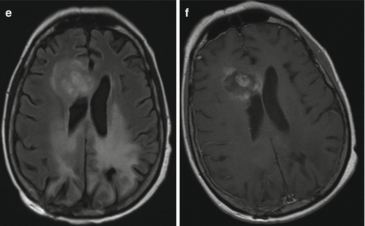
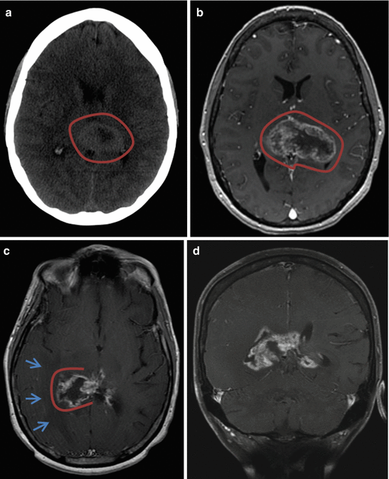
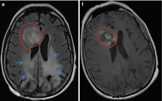
(a) and (b) are the initial CT and MRI images of the patient presenting with an intracranial mass lesion manifesting as 1 month of confusion. (c) and (d) are MRI images 8 months later showing local recurrence. (e) and (f) are MRI images from 6 months later
Questions
- 1.
Identify and describe the abnormal findings in the images shown above.
- 2.
How are brain tumors classified?
- 3.
List some of the common brain tumors.
- 4.
What is the role of steroids in the acute management of brain tumors?
- 5.
Describe important elements of anesthesia in a patient undergoing awake craniotomy for an intracranial mass lesion.
Answers
- 1.
The CT and MRI images of the brain depicted in Figs. 50.1A, B and 50.2A, B show an irregular heterogeneously enhancing mass lesion centered in the region of the splenium of the corpus callosum, slightly to the left of the midline. This mass extends to involve the left lateral ventricle and bilateral thalami. These findings are concerning for a high-grade glioma, likely glioblastoma multiforme. This patient underwent awake craniotomy and resection of the mass followed by outpatient chemotherapy and radiation. The pathology confirmed the diagnosis of glioblastoma. Figures 50.1C, D and 50.2C, D depict the local recurrence of the tumor six months later. Following this, the patient underwent repeat craniotomy and tumor resection. Figures 50.1E, F and 50.2E, F depict the pre- and post-contrast MRI images which reveal recurrence in the right frontal region eight months later (encircled region). Radiation-induced changes are also noted (blue arrows). The decision was made to pursue hospice care at this point.


Fig. 50.2


Encircled region in (a) and (b) shows the mass lesion in the region of the splenium of the corpus callosum. Open circles in (c) and (d) illustrate local recurrence, more toward the right side and the blue arrows illustrate vasogenic edema. Circled regions in (e) and (f) illustrate the recurrence in the right frontal region, while the arrows illustrate radiation-induced changes
Stay updated, free articles. Join our Telegram channel

Full access? Get Clinical Tree


