Fig. 13.1
Pathophysiology of burn shock
13.5 Classifications of Burn Injuries
13.5.1 Burn Depth
Burns are classified by the depth of tissue affected. The depth of injury depends on the degree of temperature and duration of exposure to the offending source. The deeper the burn injury, the greater the number of skin layers that are damaged. Sweat glands and hair follicle roots are located in the deeper layers and will be destroyed with a deep burn. The burn depth is usually classified as superficial (first degree), superficial-partial and deep-partial thickness (second degree), and full-thickness (third degree). Oftentimes this classification is extended to a fourth degree burn.
Superficial burn is limited to the epidermis and causes erythema and edema without vesiculation. This type of burn is painful to touch, however, it will heal without scar formation.
Superficial partial-thickness burn involves the epidermis and papillary dermis. Blister formation is present. The wounds from this type of burn typically blanch with pressure; they are painful and will heal without scar formation.
Deep partial-thickness burn involves tissue destruction up to the reticular dermis. This type of burn appears poorly vascularized and is less painful. Healing of deep partial-thickness burns occurs with severe scarring.
Full-thickness burns involve destruction of the epidermis and all layers of the dermis. This type of burn appears waxy white in color, dry, leathery, and insensate because the skin nerve endings have been destroyed. Fourth degree burns extend deeply into the subcutaneous tissue and can involve the tendon, fascia, muscle, and bone. These areas can be charred black.
Figure 13.2 illustrates clinical examples and characteristics of the burn depth classification.
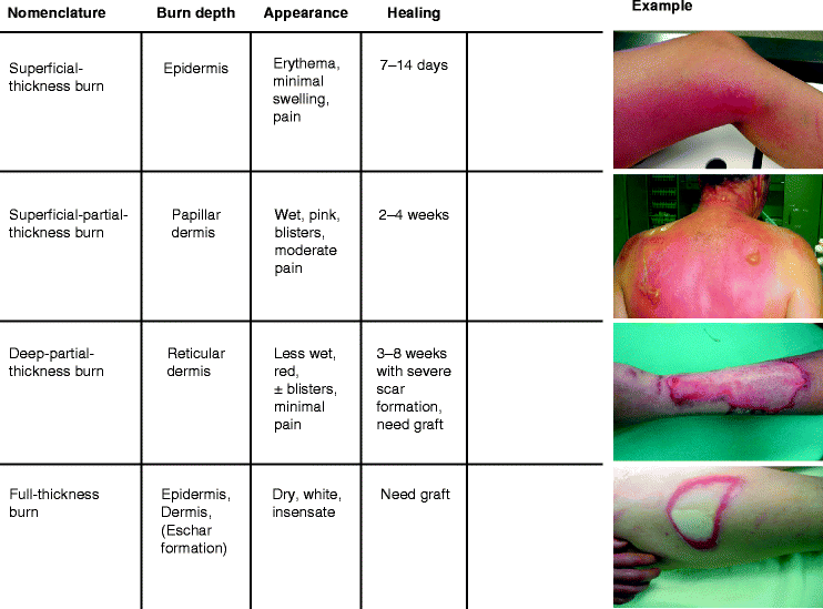
Fig. 13.2
Characteristics of superficial thickness to full-thickness burns
13.5.2 Burn Surface Area
Burns can also be classified in terms of total body surface area (TBSA). The Wallace “Rule of nines” is commonly used to determine the injured TBSA, although the measurement is adjusted for infants and children (Lund and Browder chart). The body is divided into anatomical regions that represent 9 % of the total body surface (Fig. 13.3). The outstretched palm and fingers equal 1 % of the TBSA (“Rule of palms”). Superficial burns should be excluded from the calculation.
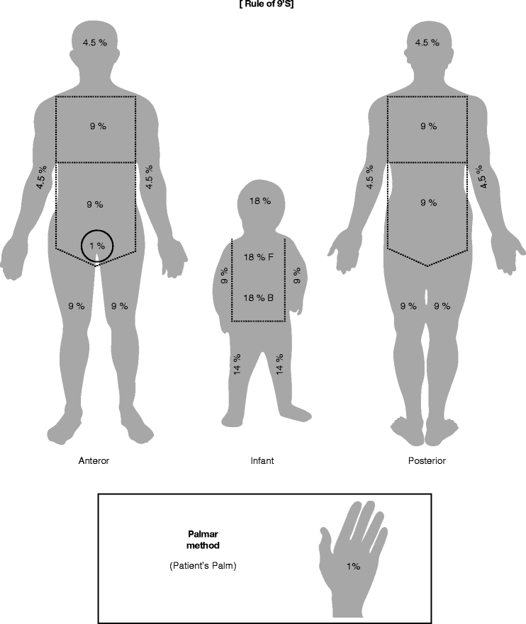
Fig. 13.3
Rules of nine to determine the body surface area
13.5.3 Transfer Criteria to a Burn Center
In order to determine the need for referral to a specialized burn unit, the German Burn Association devised the following criteria:
http://www.verbrennungsmedizin.de/leitlinien_2.htm
Burns involving specific anatomical zones (face, hands, axilla, feet, or perineum)
Burns in infants (less than 8 years old) or the elderly (older than 60 years)
Full-thickness burns over more than 10 % of the TBSA
Partial-thickness burns over more than 15 % of the TBSA
Circumferential burns to any extremity or complicated location
Burns that cross major joints
Burns associated with inhalational injury
Burns associated with fractures or other trauma
Electrical burns
Additional pre-existing diseases
13.6 Management
Accurate estimation of burn wounds is essential in determining the type of first aid administered and the need for hospitalization. Burn treatment is in large part based on burn depth and the body surface area involved.
The following points should be considered regardless of the burn depth or cause
Shock prevention
Pain control
Reduction of the risk of infection
13.6.1 Prehospital Care
Stop the burn process
Stabilize airway: consider early intubation as edema formation can later make endotracheal intubation difficult
Large-caliber intravenous lines in peripheral veins
Prevent hypothermia
Ensure adequate analgesia
Estimate the burn depth and TBSA burned
Assess the presence of other injuries (inhalation injury, fractures, visceral injuries, and lacerations)
Check transfer criteria to a burn center or other appropriate care center as indicated
13.6.2 Resuscitation
Initiation of increased fluid resuscitation in prehospital care is generally unnecessary. Clinical circulation therapy is goal-directed and takes into consideration the hemoglobin concentration, hematocrit, mean arterial pressure (MAP), diuresis, central venous pressure (CVP), and central venous sO2 [5]. Fluid resuscitation is usually necessary in burns involving more than 20 % of the TBSA in adults and more than 10 % TBSA in pediatric patients. For initial volume replacement, balanced crystalloid solutions are preferred. Colloids, gelatine solution, and catecholamines should be avoided if possible [5].
The most widely used adult formulas are the Parkland (Baxter) formula and the modified Brooke formula.

Modified Brooke formula:

Half of the fluid should be administered over the first 8 h. All resuscitation formulas are meant to serve only as guides.
13.6.3 Aspects of Burn Care
Early necrosectomy, wound closure, prevention of septic complications, adequate nutrition, and control of the external environment are of utmost importance in the management of patients with severe thermal injuries. Immediate escharotomy is indicated in deep circumferential burns of the limbs, torso, or neck to avoid compartment syndrome, respiratory distress, or reduced circulation to the limbs as a result of constriction.
Accurate burn depth determination is important, particularly differentiation of superficial-partial thickness in contrast to deep-partial thickness burns because superficial-partial thickness burns can be treated conservatively, while deep-partial thickness burns need burn wound excision and skin grafting. Various methods have been applied to determine the depth of burns [7–12]. Currently, Confocal-laser-scanning microscopy is the most promising innovation for differentiating superficial-partial and deep-partial thickness burns on a histomorphological level [13, 14].
13.6.3.1 Burn Wound Care of Superficial and Superficial Partial-Thickness Burns
Superficial and superficial partial-thickness burns can be treated conservatively as follows:
The burned area should be cooled to reduce pain and swelling by conducting heat away from the skin surface. Hypothermia and cold injury should be avoided at all costs. Blisters should be deroofed under sterile conditions, and the wounds should be covered with an antiseptic solution and a nonadhesive wound dressing. A daily wound inspection and dressing change should be performed to prevent wound infection. Alternative dressing approaches for superficial partial-thickness burns are temporary synthetic skin substitutes that could stimulate and promote wound healing. These substitutes combine synthetic membranes and biological substances and prevent, at least, painful dressing changes [15, 16].Related posts:

Stay updated, free articles. Join our Telegram channel

Full access? Get Clinical Tree


