FIGURE 22-1. Needle insertion for saphenous nerve block of the ankle.
General Considerations
An ankle block is essentially a block of four terminal branches of the sciatic nerve (deep and superficial peroneal, tibial, and sural) and one cutaneous branch of the femoral nerve (saphenous). Ankle block is simple to perform, essentially devoid of systemic complications, and highly effective for a wide variety of procedures on the foot and toes. For this reason, this technique should be in the arma mentarium of every anesthesiologist. At our institution, an ankle block is most commonly used in podiatric surgery and foot and toe debridement or amputation.
Functional Anatomy
It is useful to think of the ankle block as a block of two deep nerves (posterior tibial and deep peroneal) and three superficial nerves (saphenous, sural, and superficial peroneal). This concept is important for success of the block because the two deep nerves are anesthetized by injecting local anesthetic under the fascia, whereas the three superficial nerves are anesthetized by a simple subcutaneous injection of local anesthetic.
Common Peroneal Nerve
The common peroneal nerve separates from the tibial nerve and descends alongside the tendon of the biceps femoris muscle and around the neck of the fibula. Just below the head of the fibula, the common peroneal nerve divides into its terminal branches: the deep peroneal and superficial peroneal nerves. The peroneus longus muscle covers both nerves.
Deep Peroneal Nerve
The deep peroneal nerve runs downward below the layers of the peroneus longus, extensor digitorum longus, and extensor hallucis longus muscles to the front of the leg (Figure 22-2). At the ankle level, the nerve lies anterior to the tibia and the interosseous membrane and close to the anterior tibial artery. It is usually sandwiched between the tendons of the anterior tibial and extensor digitorum longus muscles. At this point, it divides into two terminal branches for the foot: the medial and the lateral. The medial branch passes over the dorsum of the foot, along the medial side of the dorsalis pedis artery, to the first interosseous space, where it supplies the web space between the first and second toe. The lateral branch of the deep peroneal nerve is directed anterolaterally, penetrates and innervates the extensor digitorum brevis muscle, and terminates as the second, third, and fourth dorsal interosseous nerves. These branches provide innervation to the tarsometatarsal, metatarsophalangeal, and interphalangeal joints of the lesser toes.
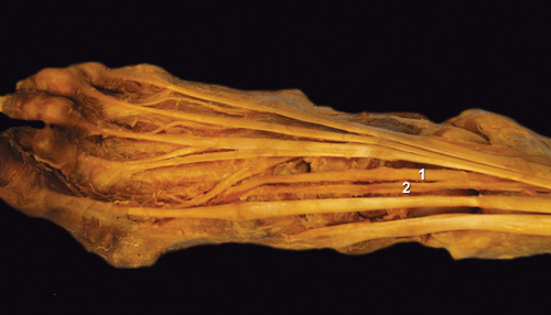
FIGURE 22-2. Anatomy of the ankle. ![]() artery dorsal pedis,
artery dorsal pedis, ![]() deep peroneal nerve.
deep peroneal nerve.
Superficial Peroneal Nerve
The superficial peroneal nerve (also called the musculocutaneous nerve of the leg) provides muscular branches to the peroneus longus and brevis muscles. After piercing the deep fascia covering the muscles, the nerve eventually emerges from the anterolateral compartment of the lower part of the leg and surfaces from beneath the fascia 5 to 10 cm above the lateral malleolus (Figure 22-3). At this point, it divides into terminal cutaneous branches: the medial and the lateral dorsal cutaneous nerves (Figure 22-4A and B). These branches carry sensory innervation to the dorsum of the foot and communicate with the saphenous nerve medially, as well as the deep peroneal nerve in the first web space and the sural nerve on the lateral aspect of the foot.
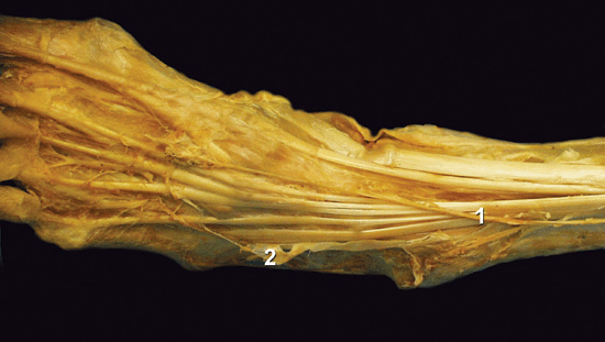
FIGURE 22-3. Anatomy of the ankle. ![]() superficial peroneal nerve,
superficial peroneal nerve, ![]() sural nerve.
sural nerve.
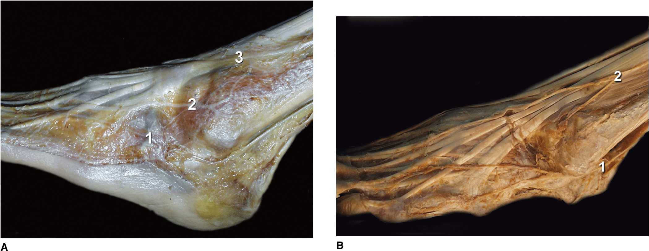
FIGURE 22-4. (A) Anatomy of the ankle. ![]() Sural nerve.
Sural nerve. ![]() ,
,![]() Superficial peroneal nerve. (B) Anatomy of the ankle. Sural nerve, Superficial peroneal nerve.
Superficial peroneal nerve. (B) Anatomy of the ankle. Sural nerve, Superficial peroneal nerve.
Tibial Nerve
The tibial nerve separates from the common popliteal nerve proximal to the popliteal fossa crease and joins the tibial artery behind the knee joint. The nerve runs distally in the thick neurovascular fascia and emerges at the inferior third of the leg from beneath the soleus and gastrocnemius muscles on the medial border of the Achilles tendon (Figure 22-5). At the level of the medial malleolus, the tibial nerve is covered by the superficial and deep fasciae of the leg. It is positioned laterally and posteriorly to the posterior tibial artery and midway between the posterior aspect of the medial malleolus and the posterior aspect of the Achilles tendon. Just beneath the malleolus, the nerve divides into lateral and medial plantar nerves. The posterior tibial nerve provides cutaneous, articular, and vascular branches to the ankle joint, medial malleolus, inner aspect of the heel, and Achilles tendon. It also branches to the skin, subcutaneous tissue, muscles, and bones of the sole.
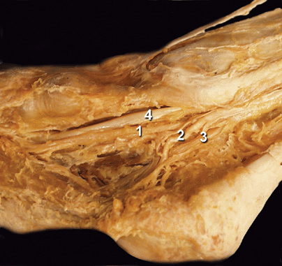
FIGURE 22-5. Anatomy of the ankle. ![]() flexor digitorum longus tendon,
flexor digitorum longus tendon, ![]() posterior tibial artery,
posterior tibial artery, ![]() tibial nerve,
tibial nerve, ![]() flexor hallucis longus.
flexor hallucis longus.
Sural Nerve
The sural nerve is a sensory nerve formed by a union of the medial sural nerve (a branch of the tibial nerve) and the lateral sural nerve (a branch of the common peroneal nerve). The sural nerve courses between the heads of the gastrocnemius muscle, and after piercing the fascia covering the muscles, it emerges on the lateral aspect of the Achilles tendon 10 to 15 cm above the lateral malleolus (Figure 22-4A and B). After providing lateral calcaneal branches to the heel, the sural nerve descends behind the lateral malleolus, supplying the lateral malleolus, Achilles tendon, and ankle joint. The sural nerve continues on the lateral aspect of the foot, innervating the skin, subcutaneous tissue, fourth interosseous space, and fifth toe.
Saphenous Nerve
The saphenous nerve is a terminal cutaneous branch (or branches) of the femoral nerve. Its course is in the subcutaneous tissue of the skin on the medial aspect of the ankle and foot (Figure 22-6).
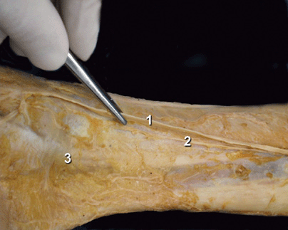
FIGURE 22-6. Anatomy of the ankle. ![]() ,
, ![]() saphenous nerve,
saphenous nerve, ![]() medial malleolus.
medial malleolus.
Related posts:


Stay updated, free articles. Join our Telegram channel

Full access? Get Clinical Tree


